. 2016 Aug 11;536(7615):224-8.
doi: 10.1038/nature19309. Epub 2016 Aug 3.

Cotranslational signal-independent SRP preloading during membrane targeting

Cotranslational signal-independent SRP preloading during membrane targeting

Justin W Chartron et al. Nature. .

Abstract

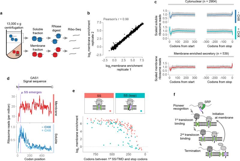
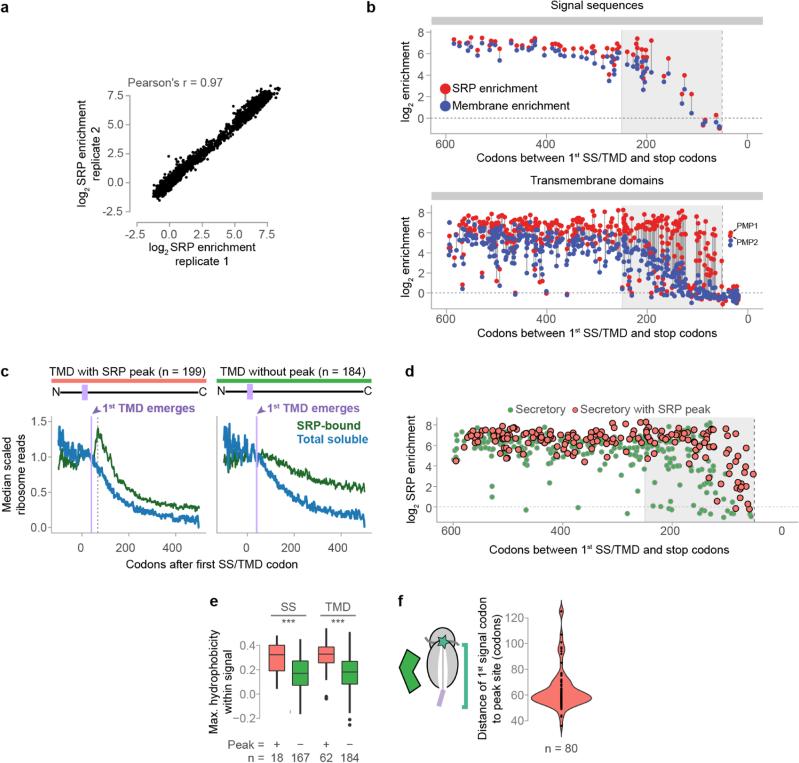
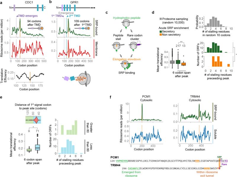
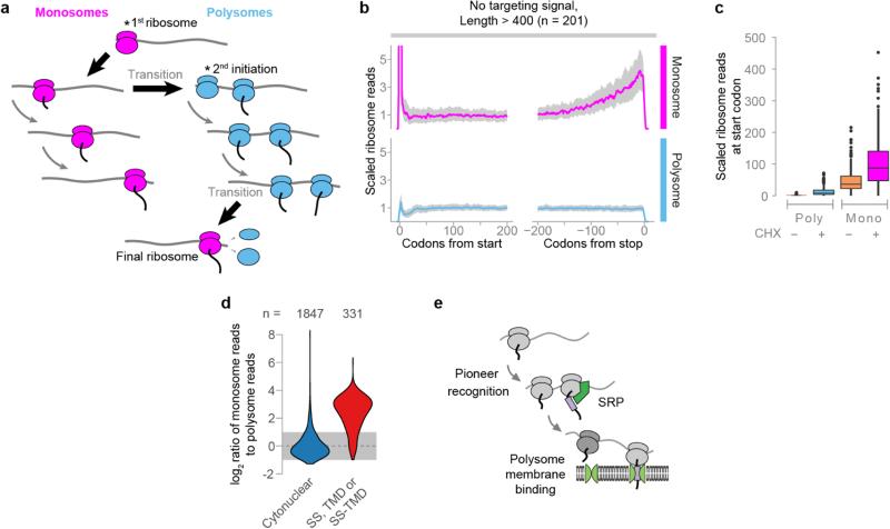
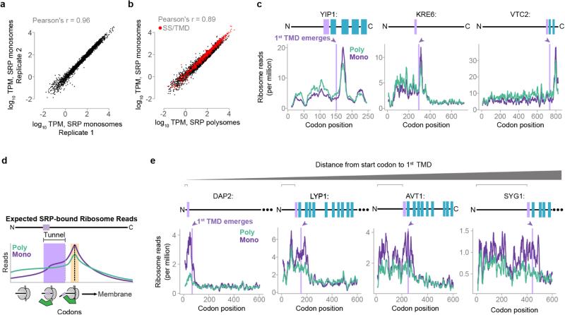
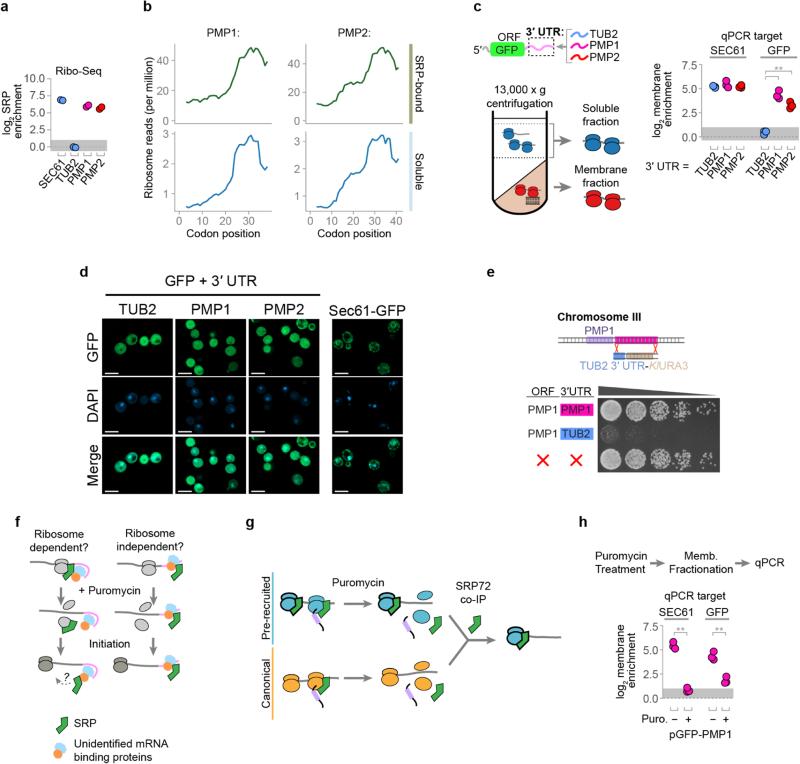
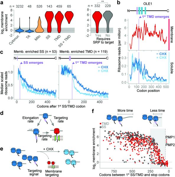
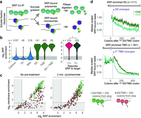
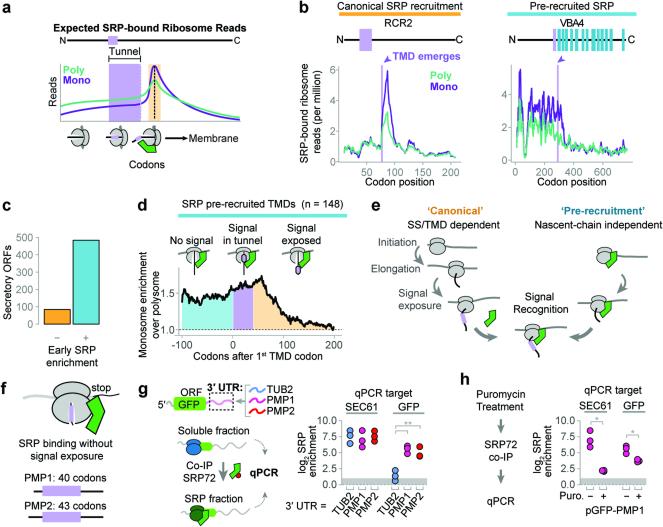
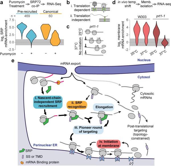
Ribosome-associated factors must properly decode the limited information available in nascent polypeptides to direct them to their correct cellular fate. It is unclear how the low complexity information exposed by the nascent chain suffices for accurate recognition by the many factors competing for the limited surface near the ribosomal exit site. Questions remain even for the well-studied cotranslational targeting cycle to the endoplasmic reticulum, involving recognition of linear hydrophobic signal sequences or transmembrane domains by the signal recognition particle (SRP). Notably, the SRP has low abundance relative to the large number of ribosome-nascent-chain complexes (RNCs), yet it accurately selects those destined for the endoplasmic reticulum. Despite their overlapping specificities, the SRP and the cotranslationally acting Hsp70 display precise mutually exclusive selectivity in vivo for their cognate RNCs. To understand cotranslational nascent chain recognition in vivo, here we investigate the cotranslational membrane-targeting cycle using ribosome profiling in yeast cells coupled with biochemical fractionation of ribosome populations. We show that the SRP preferentially binds secretory RNCs before their targeting signals are translated. Non-coding mRNA elements can promote this signal-independent pre-recruitment of SRP. Our study defines the complex kinetic interaction between elongation in the cytosol and determinants in the polypeptide and mRNA that modulate SRP–substrate selection and membrane targeting.


Figures

Extended Data Figure 1
Extended Data Figure 1. Cotranslational membrane enrichment
a, Crude lysates were fractionated, and then polysomes were recovered by sucrose gradient ultracentrifugation and used for ribosome profiling. b, Enrichment of ribosome-protected mRNA reads in the membrane polysome fractions over the soluble polysome fractions from two biological replicates. Every dot represents one ORF. c, Metagene plots of soluble polysome ribosome-protected reads of transcripts encoding proteins lacking ER targeting signals (top), or of membrane-bound polysome protected reads of transcripts encoding secretory proteins that were at least 2-fold membrane-enriched (bottom). For each ORF, ribosome-protected reads at each position were scaled by dividing by the mean reads per codon of the ORF, excluding first two and last two sense codons. The median scaled reads at each position are plotted as a line, and the interquartile range is shaded in gray. d, Ribosome-protected reads at each codon of an example secreted protein, β-1,3-glucanosyltransferase (GAS1), a model SRP-independent protein. Topology is indicated above, with the signal sequence in lavender. The position where the signal begins to emerge from the ribosome exit tunnel is indicated. e, The number of codons remaining after the encoding of the first residue of a SS, and the corresponding membrane enrichment per SS containing ORF. Signal sequences were divided between those that bind Ssh1p directly upon exposure and those that require a looped conformation (>90 codons after the first SS codon) . f, Transcripts remain at the membrane by subsequent translocon binding, thus the small soluble fraction comprises mRNA undergoing initial targeting.
Extended Data Figure 2
Extended Data Figure 2. Cotranslational enrichment of SRP
a, Enrichment of ribosome-protected mRNA reads in the soluble SRP-bound polysome fractions over the total soluble polysome fractions from two biological replicates. b, The number of codons remaining after encoding of first SS or TMD residue, and the corresponding SRP and membrane enrichment scores per ORF. Scores are determined from cultures harvested without added cycloheximide. Enrichment scores are indicated with filled dots, and the scores from the same transcript are linked with a gray line. The vertical dashed line indicates 50 codons, the boundary for TA proteins. Here, only SS that bind Ssh1p directly upon exposure from the RNC are shown. c, Secretory transcripts were classified into two groups based on the ribosome protected-read distributions from SRP-bound polysomes. Some displayed a pronounced increase of reads at positions coincident with to the initial exposure of and SS or TMD by the ribosome, while others did not. Shown here are metagene analysis plots of soluble polysome protected reads from the categorized TMD proteins. For each ORF, the reads at each codon position were divided by the mean reads per codon within the range +20 to +40 after first signal codon. The first 30 codons of each ORF are excluded to avoid the characteristic low-density region near the start codon. The lavender line indicates when the first TMD begins to emerge from the exit tunnel, and the dashed line indicates the position of the read peak. Notably, the total soluble polysome reads depleted in a similar manner for both classes, a read increase was not observed in the total soluble reads, and reads from the SRP-bound transcripts with a peak did not deplete faster than the total soluble reads. These features are consistent with a model where SRP is recruited at the peak site, and elongation then proceeds at the same rate. d, The number of codons remaining after encoding of first SS or TMD and corresponding SRP enrichment. Transcripts are classified by the presence or absence of a read increase following signal exposure, as in c. Note that for SRP-enriched transcripts with signals closest to the terminus (<100 codons), evidence of direct binding between SRP and the nascent chain was always observed. SRP can therefore bind late transmembrane domains immediately after they become exposed by the ribosome. e, Maximum hydrophobicity across targeting signals using an 8-residue averaging window. Only signals with peaks that could be unambiguously attributed to a targeting signal were included. Hydrophobicity was determined by attributing the biological hydrophobicity score to each encoded amino acid. ***p ≤ 0.001, Wilcoxon rank-sum test. f, Distribution of the distance between the first codon of a targeting signal and the position of the downstream read increase. Only transcripts wherein the increase can be unambiguously attributed to a specific targeting signal were included.
Extended Data Figure 3
Extended Data Figure 3. Elongation pausing and local SRP recruitment
a, b, Local increases of ribosome-protected reads from membrane-bound polysomes, indicated with orange lines, were coincident with rare codons, as in cell division cycle protein 1 (CDC1, a) or polybasic nascent chains, as in the plasma membrane G-protein coupled receptor (GPR1, b). Soluble SRP-bound polysome-protected reads were further increased at the same positions. c, In these cases, hydrophobic sequences in the nascent chain were exposed to the cytosol at the locations of increased reads, which were coincident with elongation attenuators. d, Translational efficiencies for the 6 codons following, and the number of stalling residues within the 10 residues preceding, the sites of increased SRP-bound ribosome reads. Translational efficiency was determined by attributing the nTE score to each codon. Residues that were found to stall the ribosome, based on previous investigation,,, were lysine, arginine, glutamate, aspartate, proline and glycine. Because of variation in specific motifs, and uncertainty in whether these motifs are additive, we simply compared the total number of these residues in the indicated 10 residue spans. Sets of 10,000 random sequences, at least 10 amino acids from the stop codon, were sampled from 5907 non-dubious ORFs, and translational efficiency and stalling residues were determined over 6 or 10 codon spans. *p ≤ 0.05, **p ≤ 0.01, Wilcoxon rank-sum tests. e, The targeting signals that recruited SRP directly to the nascent chain unusually far from the encoding of the signal had SRP-binding sites coincident with intrinsic elongation attenuation. Secretory protein transcripts that showed an increase in SRP-bound protected reads (see Extended Data Fig. 2c, f) were further classified by the position of the peak relative to the first signal codon. Transcripts with peaks found at least 80 codons after the signal had significantly lower translational efficiency in the 6 codons following the peak. These transcripts also had a greater, but not statistically significant, amount of stalling amino acids in the 10 residues preceding the peak. *p ≤ 0.05, Wilcoxon rank-sum tests. f, Similar increases in SRP-bound reads were observed for certain non-secretory proteins as exemplified by phosphoacetylglucosamine mutase (PCM1) and tRNASer Um44 2′-O-methyltransferase (TRM44). Hydrophobic sequences in non-secretory proteins, coupled with attenuation of elongation, may lead to SRP recruitment.
Extended Data Figure 4
Extended Data Figure 4. Ribosome profiling of monosomes
a, Ribosomes transition from monosomes to polysomes during elongation. The pioneer round of initiation will be a monosome, and during elongation there is a chance of additional initiation converting the transcript to a polysome. Similarly, a polysome will become a monosome if all ribosomes but one terminate. As mRNA is sampled closer to the stop codon, the likelihood of observing a footprint from the final ribosome will increase. b, Metagene analysis of soluble monosome or polysome protected reads from proteins lacking an ER targeting signal. Data were obtained using cycloheximide treatment. ORFs are at least 400 codons long and have an average of at least 0.5 reads per codon in each dataset. For each ORF, ribosome reads at each position were divided by the mean reads per codon over the range +160 to +240 codons. The median normalized read value at each codon position is plotted, and the interquartile range is shaded in gray. c, Relative reads at the start codon from ORFs normalized in b. d, Distributions of the ratio of ribosome-protected reads found in soluble monosomes over soluble polysomes. e, A pioneer round of translation deposits mRNA on the membrane. Polysomes will be retained at the membrane and are therefore depleted from the soluble fraction.
Extended Data Figure 5
Extended Data Figure 5. Ribosome profiling of SRP-bound monosomes
a, Ribosome-protected reads, in tags per million (TPM) for each ORF, from SRP-bound monosome fractions from two biological replicates. b, Ribosome-protected reads from the soluble SRP-bound monosome and SRP-bound polysome fractions of the same biological replicate, with cycloheximide treatment. c, Distribution of ribosome reads within example ORFs that display SRP-bound monosome and polysome profiles consistent with direct recognition of the nascent chain. d, If RNCs can recruit SRP while a TMD is within the exit tunnel, then there will be an increase in ribosome-protected reads from SRP-bound monosomes when the TMD begins to translate (lavender). This increase will maximize when the TMD is exposed to the cytosol (orange). e, Distribution of ribosome reads within example ORFs that display SRP-bound monosome profiles consistent with recruitment to transcripts prior to targeting signal synthesis. Examples are arranged for an increasing distance from the start codon to the first TMD. Only the first 600 codons for each ORF are shown.
Extended Data Figure 6
Extended Data Figure 6. The role of the UTR from PMP1 and PMP2
a, The cotranslational SRP enrichment of the PMP1 and PMP2 ORFs was similar to other bona fide secretory proteins, such as SEC61. In contrast, cytosolic proteins such as tubulin (TUB2), were not enriched. The enrichment scores are determined from the SRP-bound and total soluble polysomes from two biological replicates harvested without added cycloheximide. b, Distribution of ribosome-protected reads from soluble polysomes within the PMP1 and PMP2 ORFs. c, Membrane enrichment, determined by qPCR, of the mRNA of GFP fused to the indicated 3′ UTRs. The coding sequence of endogenous SEC61 transcript was also amplified as a control for a membrane localized transcript. **p ≤ 0.01, n = 3 biological replicates, Welch's t-test. d, Localization of mature GFP. The scale bar indicates 5 μM. Yeast were grown to mid-log phase and imaged using an Axio Observer Z1 with a Plan-Apochromat 100x/1.4 oil immersion objective (Zeiss). Z-stacks were deconvoluted by the iterative maximum likelihood algorithm in ZEN (Zeiss) and single planes are shown. Images were representative from a set of 2 replicated assays. e, Yeast growth after replacement of the endogenous 3′ UTR of PMP1 with the 3′ UTR of tubulin. Also shown is a complete deletion of PMP1 ORF. Gibson assembly was used to fuse the 300 nt TUB2 3′ UTR to the KlURA3 cassette into SmaI digested pUC19. The TUB2-UTR-URA3 element was PCR amplified, including 40 nt overhangs matching genomic sequences, and replaced the 650 nt immediately following the PMP1 coding sequence in strain BY4741 by homologous recombination. Image is representative from a set of 3 replicated assays. f, Nascent chain independent SRP recognition may require ribosomes. Puromycin treatment of lysates disrupts elongating, but not initiating, ribosomes. g, Transcripts showing only canonical recognition are more sensitive to puromycin. This is consistent with puromycin resistance of SRP that has pre-recruited to initiating ribosomes. h, Membrane enrichment of the GFP-PMP1 construct or SEC61 mRNA after lysates were incubated with puromycin. **p ≤ 0.01, n = 3 biological replicates, Welch's t-test.
Extended Data Figure 7
Extended Data Figure 7. The role translation in membrane enrichment
a, Lysates were treated with puromycin prior to membrane fractionation. mRNA recovered from the soluble and membrane fractions were used for RNA-seq b, Membrane enrichment of secretory protein transcripts (SS, TMD, SS-TMD, or TA, n = 729) following puromycin treatment of lysates.
Figure 1
Figure 1. Cotranslational membrane enrichment
a, Distributions of the open reading frame (ORF) enrichment of ribosome-protected reads in the membrane fraction over the soluble fraction. ORFs were alternatively classified by expected SRP dependence. **p ≤ 0.01, Wilcoxon rank-sum test. b, Ribosome-protected reads at each codon of an example transmembrane protein OLE1. Membrane topology is indicated above, with the first TMD in lavender. c, Metagene analysis of soluble fraction polysome-protected reads from transcripts that were at least 2-fold membrane enriched. ORFs were aligned at the targeting signal and scaled. d, Cotranslational membrane targeting is in competition with elongation. e, Elongation inhibitors provide additional time for polysomes exposing a targeting signal to localize to the membrane. f, Membrane enrichment was limited by the length of the reading frame remaining after the encoding of targeting signals. The vertical dashed line indicates 50 codons.
Figure 2
Figure 2. Cotranslational enrichment of SRP
a, Srp72p-TAP was immunoprecipitated from the total soluble fraction. SRP-bound monosomes and polysomes were separated by sucrose gradient ultracentrifugation. b, Distributions of the ORF enrichment of ribosome-protected reads from SRP-bound soluble polysomes over the total soluble polysomes. ORFs were alternatively classified by expected SRP dependence. *p ≤ 0.05, Wilcoxon rank-sum test. c, Cotranslational membrane-fraction enrichment compared to SRP enrichment. d, Metagene analysis of soluble SRP-bound polysome-protected reads from transcripts that are at least 2-fold SRP-enriched. ORFs were aligned at the targeting signal and scaled.
Figure 3
Figure 3. Distinct mechanisms of SRP recruitment
a, Recruitment of SRP to RNCs is expected to increase ribosome-protected reads from SRP-bound monosomes when an SS or TMD is exposed to the cytosol (orange). b, Distributions of SRP-bound ribosome reads on representative transcripts from cycloheximide-treated cultures. Selected transcripts are RCR2, and VBA4. c, The majority of secretory proteins demonstrated SRP enrichment prior to signal exposure. d, Metagene plot of the median value of enrichment of SRP-bound monosomes over polysomes. Included transcripts encode TMDs at least 40 codons from the start codon. Shaded areas represent enrichment before the TMD is encoded (cyan), while the TMD is in the ribosome exit tunnel (lavender), and after the TMD is exposed (orange). e, Two mechanisms for SRP to select secretory mRNA. f, PMP1 and PMP2 were the only TA proteins that enriched SRP. g, The GFP ORF was fused to the indicated 3′ UTRs and expressed in vivo. Srp72p-TAP was immunoprecipitated from the total soluble fraction and RNAs were subject to qPCR. **p ≤ 0.01, n = 3 biological replicates, Welch's t-test. h, Puromycin treatment of lysate from yeast expressing GFP with the PMP1 3′ UTR was followed by SRP immunoprecipitation and qPCR. *p ≤ 0.05, n = 3 biological replicates, Welch's t-test.
Figure 4
Figure 4. Translation and the role of SRP
a, Distributions of RNA-seq SRP enrichment scores from secretory protein transcripts (SS, TMD, SS-TMD, or TA), with or without puromycin treatment. Included ORFs have at least 2-fold SRP enrichment without puromycin. b, The prt1-1 allele prevents initiation at non-permissive temperatures. Translational run-off removes all ribosomes from transcripts. b, Transcripts are retained on the membrane though binding of the RNC to the translocon. It is also possible that mRNA binding proteins at the ER bind transcripts. c, Distributions of RNA-seq membrane enrichment scores of secretory protein transcripts (n = 584). d, After mRNA export, a pioneer round of targeting directs secretory transcripts to the ER membrane. SRP is specifically pre-recruited transcripts that will present a functional targeting signal. Upon emergence of an SS or TMD, SRP directs RNCs to the ER membrane. Once at the ER membrane, transcripts are retained over multiple rounds of translation.

References

    1. Pechmann S, Willmund F, Frydman J. The ribosome as a hub for protein quality control. Mol Cell. 2013;49:411–421. - PMC - PubMed
    1. Bornemann T, Holtkamp W, Wintermeyer W. Interplay between trigger factor and other protein biogenesis factors on the ribosome. Nat Commun. 2014;5:4180. - PubMed
    1. Nyathi Y, Pool MR. Analysis of the interplay of protein biogenesis factors at the ribosome exit site reveals new role for NAC. J Cell Biol. 2015;210:287–301. - PMC - PubMed
    1. Akopian D, Shen K, Zhang X, Shan SO. Signal recognition particle: an essential protein-targeting machine. Annu Rev Biochem. 2013;82:693–721. - PMC - PubMed
    1. Zhang X, Shan SO. Fidelity of cotranslational protein targeting by the signal recognition particle. Annu Rev Biophys. 2014;43:381–408. - PMC - PubMed

Methods Online References

    1. Ghaemmaghami S, et al. Global analysis of protein expression in yeast. Nature. 2003;425:737–741. - PubMed
    1. Huh WK, et al. Global analysis of protein localization in budding yeast. Nature. 2003;425:686–691. - PubMed
    1. Zhong T, Arndt KT. The yeast SIS1 protein, a DnaJ homolog, is required for the initiation of translation. Cell. 1993;73:1175–1186. - PubMed
    1. Winzeler EA, et al. Functional characterization of the S. cerevisiae genome by gene deletion and parallel analysis. Science. 1999;285:901–906. - PubMed
    1. Freidlin PJ, Patterson RJ. Heparin releases monosomes and polysomes from rough endoplasmic reticulum. Biochem Biophys Res Commun. 1980;93:521–527. - PubMed

Publication types

MeSH terms