. 2017 Feb;28(2):452-459.
doi: 10.1681/ASN.2016020232. Epub 2016 Aug 3.

Fully Automated Evaluation of Total Glomerular Number and Capillary Tuft Size in Nephritic Kidneys Using Lightsheet Microscopy

Affiliations

Fully Automated Evaluation of Total Glomerular Number and Capillary Tuft Size in Nephritic Kidneys Using Lightsheet Microscopy

Anika Klingberg et al. J Am Soc Nephrol. 2017 Feb.

Abstract

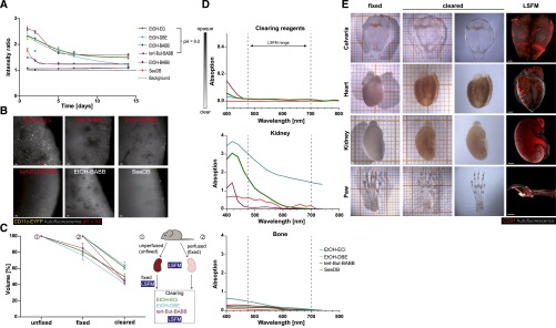
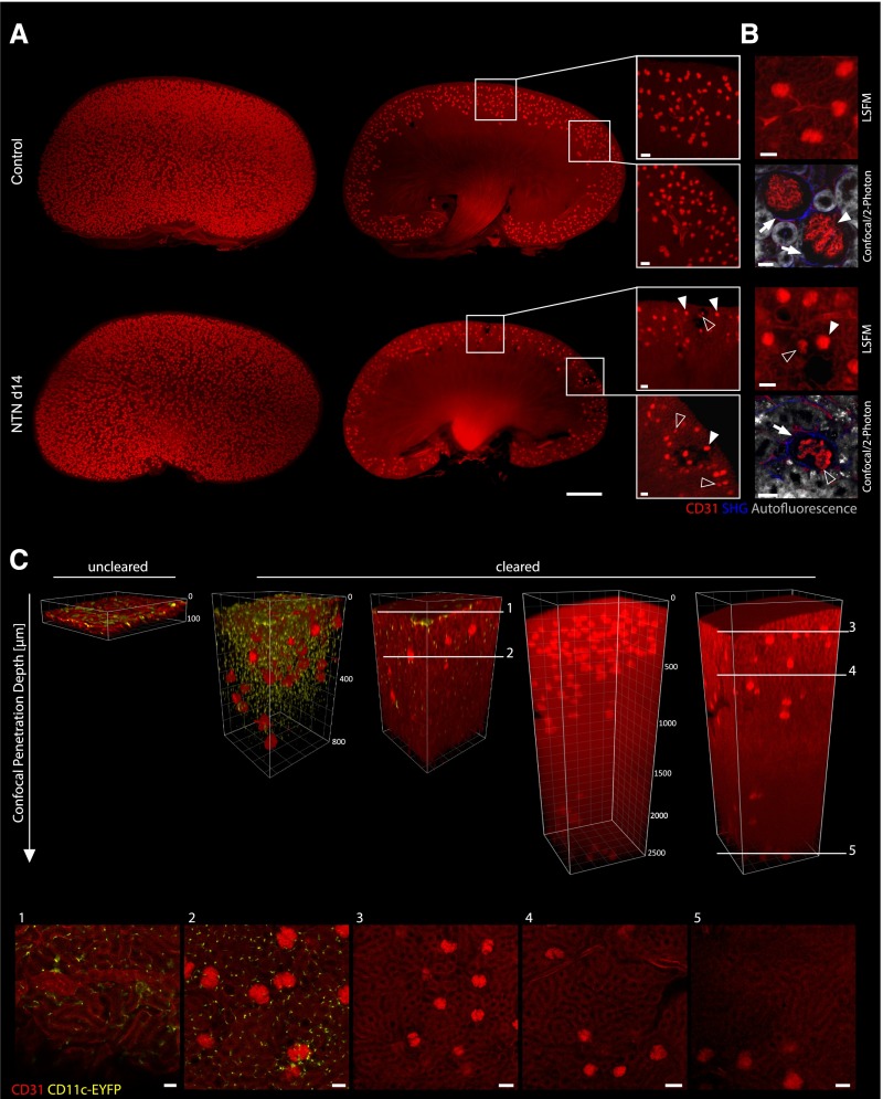
The total number of glomeruli is a fundamental parameter of kidney function but very difficult to determine using standard methodology. Here, we counted all individual glomeruli in murine kidneys and sized the capillary tufts by combining in vivo fluorescence labeling of endothelial cells, a novel tissue-clearing technique, lightsheet microscopy, and automated registration by image analysis. Total hands-on time per organ was <1 hour, and automated counting/sizing was finished in <3 hours. We also investigated the novel use of ethyl-3-phenylprop-2-enoate (ethyl cinnamate) as a nontoxic solvent-based clearing reagent that can be handled without specific safety measures. Ethyl cinnamate rapidly cleared all tested organs, including calcified bone, but the fluorescence of proteins and immunohistochemical labels was maintained over weeks. Using ethyl cinnamate-cleared kidneys, we also quantified the average creatinine clearance rate per glomerulus. This parameter decreased in the first week of experimental nephrotoxic nephritis, whereas reduction in glomerular numbers occurred much later. Our approach delivers fundamental parameters of renal function, and because of its ease of use and speed, it is suitable for high-throughput analysis and could greatly facilitate studies of the effect of kidney diseases on whole-organ physiology.

Keywords: Immunology and pathology; glomerular endothelial cells; glomerular filtration rate; glomerulonephritis; immune complexes; kidney anatomy.


Figures

Figure 1.
Figure 1.
Optical clearing via Ethanol-ECi allows clearing of soft and hard tissues with prolonged fluorescence maintenance. (A) Long–term EYFP fluorescence over background in kidneys of CD11c-EYFP mice after clearing with different protocols. pH values of dehydration reagents were adjusted where indicated. Data are means±SEM of 100 measured cell-to-background ratios from one kidney per condition measured repetitively at all time points. (B) Representative images from kidneys at day 14 after clearing with the indicated protocols. Red text indicates pH adjustment of dehydration reagent. Scale bars, 50 μm. (C) Volume shrinkage of kidneys induced by perfusion/fixation and different clearing protocols measured from LSFM data using automated image analysis. The schematic of the experiment is shown on the right. Data are means±SEM of three kidneys each analyzed repetitively per condition. From each animal, one kidney was unperfused, and one was perfused. In total, nine animals (18 kidneys) were analyzed. (D) Transparency of clearing reagents, kidney, or bone after clearing with the indicated protocols measured spectrophotometrically. The wavelength range used for excitation/emission in LSFM is indicated with dashed lines. Data are means from five technical replicates of one sample measured per condition. SEMs are not shown because of extremely small variations of data. (E) Optical clearing via ECi is working for soft tissues (kidney and heart) and hard tissues (calvarial or long bones). In addition to EYFP preservation, fluorescence labeling by antibodies resists the clearing procedure as shown via endothelial specific staining (CD31; red). Paw =100 μm. Grid size (in left two columns) is 1 mm (small squares). Data are representative of 10 organs imaged similarly. EtOH, ethanol. Scale bars, 1000 μm.
Figure 2.
Figure 2.
Visualization of 3D vessel trees and dendritic cells in entire ECi-cleared kidneys via LSFM and confocal/two-photon imaging. LSFM of specifically stained endothelial structures (CD31; red) allows three-dimensional reconstruction of whole kidneys. (A) Three-dimensional reconstructions of kidneys from a healthy control or an organ suffering from NTN (day 14). Note that the NTN kidney shows lower glomerular density, whereas two–dimensional optical sections reveal CD31-negative areas of corrupted glomeruli and the surrounding vasculature compared with a homogeneous field of equally sized glomeruli in healthy controls (white boxes). Furthermore, a higher heterogeneity of glomerular tuft sizes was observable in NTN kidneys at day 14. Many shrunken glomerular tufts (open arrowheads) were visible next to normally sized elements (white arrowheads). Scale bars, 50 μm. (B) Enhanced views of kidney structure (autofluorescence [gray] and fibrous tissue [second harmonic generation (SHG); blue]) and glomerular tuft (CD31; red) via combined confocal and two–photon laser scanning microscopy (LSM) compared with enhanced LSFM magnification. Compared with controls, NTN kidneys showed decreased tuft size (open arrowheads; as opposed to normal tuft size [white arrowheads]), decrease of endothelial CD31 label in their capillaries, and increased tissue fibrosis (indicated by higher SHG signal; white arrows) around damaged glomeruli. Scale bars, 20 μm in confocal/two-photon microscopy; 50 μm in LSFM. (C) Penetration depth of confocal LSM into ECi-cleared kidneys from CD11c-EYFP animals is enhanced compared with in uncleared samples. The maximum penetration depth of short wavelengths was increased from approximately 50 to 800 μm as shown via detection of CD11c-eYFP+ cells. The detected CD11c-eYFP+ cells still show the characteristic dendritic morphology after ECi clearing in the entire kidney tissue as emphasized at two exemplary focal planes of the shown Z stack (1 and 2). Longer wavelengths of the used endothelial marker CD31-Alexa Fluor-647 (red) can be detected even deeper in the tissue. The objective of the used confocal microscope allows a maximum penetration depth of 2500 μm because of the working distance. This penetration depth was reached in cleared kidneys and still allowed the discrimination of glomeruli from the surrounding tubular structures as indicated with the white lines at different focal planes (3–5).
Figure 3.
Figure 3.
Fully automated quantification of functional elements in healthy and nephritic kidneys. (A) On the basis of whole-organ images, the total kidney volume was calculated by automated image analysis and shows the typical swelling of NTN kidneys at day 7 and declining kidney size down to control levels at day 14. **P<0.05 (two–tailed, unpaired Kruskal–Wallis H test); ***P<0.001 (two–tailed, unpaired Kruskal–Wallis H test). (B) The total numbers of glomeruli per kidney were quantified with a fully automated image processing algorithm showing a highly significant loss of glomeruli at day 14 of NTN compared with day 7 and controls. **P<0.05 (two–tailed, unpaired Kruskal–Wallis H test). (C) Distribution of tuft volumes of NTN mice at day 7 and day 14 relative to the distribution in healthy controls. The glomerular tuft volumes at day 14 of NTN are increasing compared with control and day 7 of NTN as indicated via their median values (dashed lines). For the generation of B and C, a total number of 302,023 glomeruli from 23 kidneys was counted, and their tufts were individually sized by voxel counting. ****P<0.001 (chi-squared test). Daily urine analysis shows loss of glomerular filtration functionality indicated by (D) increasing albumin-to-creatinine (Alb/Crea) ratio and (E) decreasing creatinine (Crea) clearance (nonsignificant two–tailed, unpaired Kruskal–Wallis H test). (F) On the basis of automated quantification of glomeruli, the creatinine clearance efficiency per glomerulus was calculated, showing the decreasing clearance efficiency at day 7 and day 14 compared with in control mice (nonsignificant two–tailed, unpaired Kruskal–Wallis H test). For D–F, data of n=2 control animals and n=3 NTN-treated animals at day 7 and day 14 were analyzed.

References

    1. Kurts C, Panzer U, Anders HJ, Rees AJ: The immune system and kidney disease: Basic concepts and clinical implications. Nat Rev Immunol 13: 738–753, 2013 - PubMed
    1. Bertram JF, Douglas-Denton RN, Diouf B, Hughson MD, Hoy WE: Human nephron number: Implications for health and disease. Pediatr Nephrol 26: 1529–1533, 2011 - PubMed
    1. Murawski IJ, Maina RW, Gupta IR: The relationship between nephron number, kidney size and body weight in two inbred mouse strains. Organogenesis 6: 189–194, 2010 - PMC - PubMed
    1. Stelloh C, Allen KP, Mattson DL, Lerch-Gaggl A, Reddy S, El-Meanawy A: Prematurity in mice leads to reduction in nephron number, hypertension, and proteinuria. Transl Res 159: 80–89, 2012 - PMC - PubMed
    1. Baldelomar EJ, Charlton JR, Beeman SC, Hann BD, Cullen-McEwen L, Pearl VM, Bertram JF, Wu T, Zhang M, Bennett KM: Phenotyping by magnetic resonance imaging nondestructively measures glomerular number and volume distribution in mice with and without nephron reduction. Kidney Int 89: 498–505, 2016 - PMC - PubMed

Publication types