. 2016 Aug;43(8):4775.
doi: 10.1118/1.4958961.

Development of bimetallic (Zn@Au) nanoparticles as potential PET-imageable radiosensitizers

Affiliations

Development of bimetallic (Zn@Au) nanoparticles as potential PET-imageable radiosensitizers

Jongmin Cho et al. Med Phys. 2016 Aug.

Abstract

Purpose: Gold nanoparticles (GNPs) are being investigated actively for various applications in cancer diagnosis and therapy. As an effort to improve the imaging of GNPs in vivo, the authors developed bimetallic hybrid Zn@Au NPs with zinc cores and gold shells, aiming to render them in vivo visibility through positron emission tomography (PET) after the proton activation of the zinc core as well as capability to induce radiosensitization through the secondary electrons produced from the gold shell when irradiated by various radiation sources.

Methods: Nearly spherical zinc NPs (∼5-nm diameter) were synthesized and then coated with a ∼4.25-nm gold layer to make Zn@Au NPs (∼13.5-nm total diameter). 28.6 mg of these Zn@Au NPs was deposited (∼100 μm thick) on a thin cellulose target and placed in an aluminum target holder and subsequently irradiated with 14.15-MeV protons from a GE PETtrace cyclotron with 5-μA current for 5 min. After irradiation, the cellulose matrix with the NPs was placed in a dose calibrator to assess the induced radioactivity. The same procedure was repeated with 8-MeV protons. Gamma ray spectroscopy using an high-purity germanium detector was conducted on a very small fraction (<1 mg) of the irradiated NPs for each proton energy. In addition to experimental measurements, Monte Carlo simulations were also performed with radioactive Zn@Au NPs and solid GNPs of the same size irradiated with 160-MeV protons and 250-kVp x-rays.

Results: The authors measured 168 μCi of activity 32 min after the end of bombardment for the 14.15-MeV proton energy sample using the (66)Ga setting on a dose calibrator; activity decreased to 2 μCi over a 24-h period. For the 8-MeV proton energy sample, PET imaging was additionally performed for 5 min after a 12-h delay. A 12-h gamma ray spectrum showed strong peaks at 511 keV (2.05 × 10(6) counts) with several other peaks of smaller magnitude for each proton energy sample. PET imaging showed strong PET signals from mostly decaying (66)Ga. The Monte Carlo results showed that radioactive Zn@Au NPs and solid GNPs provided similar characteristics in terms of their secondary electron spectra when irradiated.

Conclusions: The Zn@Au NPs developed in this investigation have the potential to be used as PET-imageable radiosensitizers for radiotherapy applications as well as PET tracers for molecular imaging applications.


Figures

FIG. 1.
FIG. 1.
(a) Configuration of synthesized gold-coated zinc (Zn@Au) NPs. This model assumed a nominal diameter/thickness (5/4.25 nm) of zinc core and gold shell. (b) When irradiated with protons, the zinc core is activated to generate positron emitters (64Ga, 66Ga, and 68Ga). Note the volume and mass fractions (in %) of Au and Zn in each Zn@Au NP are 94.9% and 5.1%, and 98.1% and 1.9%, respectively.
FIG. 2.
FIG. 2.
Nuclear interaction cross sections leading to radioactive gamma ray, positron, and electron emissions for natural zinc and gold bombarded with low-energy protons (data obtained from Brookhaven National Laboratory EXFOR library). When bombarded with protons, 68Zn, 66Zn, and 64Zn are activated to create 68Ga, 66Ga, and 64Ga, which decay via positron emission with high-branching ratios (B+) of 90%, 57%, and 98%. The natural abundance of 68Zn is 18.8%; for 66Zn, 27.9%; and for 64Zn, 48.6%. One hundred percent of natural gold exists as 197Au. Two vertical lines represent the proton energies (14.15 and 8 MeV) used to bombard gold-coated zinc (Zn@Au) NPs. The nuclear interaction energy threshold of gold occurs at ∼8 MeV.
FIG. 3.
FIG. 3.
Setup for the second Monte Carlo simulations. The incident proton or x-ray beam interacted with nonradioactive gold-coated zinc (Zn@Au) NPs or GNPs placed in vacuum, as well as with the secondary particles. All secondary particles scored at the surface of NP.
FIG. 4.
FIG. 4.
Setup for the third Monte Carlo simulations. 66Ga atoms that decay with positron, electron, and photon emission were randomly distributed inside the Zn core of a Zn@Au NP. The source photons and electrons from decaying 66Ga radioisotopes were anticipated to attenuate and degrade in energy while escaping the Zn@Au NP.
FIG. 5.
FIG. 5.
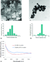
Transmission electron microscopy images and particle diameter histograms of (a) zinc NPs and (b) gold-coated zinc (Zn@Au) NPs. (c) UV-vis spectra of Zn NPs and Zn@Au NPs in water.
FIG. 6.
FIG. 6.
Time activity curve (measured activity) of gold-coated zinc (Zn@Au) NPs bombarded with 14.15-MeV protons. The curve was least-squares fitted with 3 simple exponential decay curves of 66Ga (T½ = 9.50 h), 68Ga (T½ = 67.72 min), and 13N (T½ = 9.97 min).
FIG. 7.
FIG. 7.
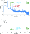
Gamma ray spectrum acquired for 12 h for gold-coated zinc (Zn@Au) NP samples bombarded with 14.15-MeV protons. (a) Gamma ray counting started 2 h after EOB (for a < 1 mg sample). A total of 2.05 × 10⁶ counts of 511-keV gamma rays were recorded for the full-width half-maximum of the peak over 511 ± 1.5 keV. (b) Gamma ray counting started 70 h after EOB (for a 28.6-mg sample).
FIG. 8.
FIG. 8.
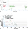
Gamma ray spectrum acquired for 1.5 h for a gold-coated zinc (Zn@Au) NP sample (<1 mg) bombarded with 8-MeV protons. Gamma ray counting started 4.5 h after EOB. (a) Logarithmic scale. All solid (green) arrows are gamma ray spectral lines from 66Ga. (b) Linear scale. A total of 680 000 counts of 511-keV gamma rays were recorded for the full-width half-maximum of the peak over 511 ± 1.5 keV.
FIG. 9.
FIG. 9.
Computed tomography (CT; left), positron emission tomography (PET; middle), and PET/CT fusion (right) images of the 23-mg gold-coated zinc (Zn@Au) NP sample irradiated with 8-MeV protons. The PET scan was started 12 h after EOB and acquired for 5 min.
FIG. 10.
FIG. 10.
Monte Carlo-simulated secondary electrons scored at the surface of each NP. (a) NPs were bombarded with 160-MeV protons at the center of modulation. (b) NPs were bombarded with 250-kVp x-rays. GNP: gold nanoparticle. Zn@Au NP: gold-coated zinc nanoparticle.
FIG. 11.
FIG. 11.
Energy spectra of the source photons and electrons (with branching ratio >0.3%) of decaying 66Ga (normalized per disintegration which emits 2.2 photons and 0.8 electrons per 66Ga decay) and the changes in the energy spectra after escaping Zn@Au NP. (a) There were negligible attenuation and energy degradation for photons (blue spectral peaks are nearly identical to red spectral peaks in the figure). The percentage of secondary photons was nearly zero. (b) Moderate self-absorption (17%) and energy degradation of primary electrons were noted as shown while escaping Zn@Au NPs. (See color online version.)

References

    1. Brun E., Sanche L., and Sicard-Roselli C., “Parameters governing gold nanoparticle x-ray radiosensitization of DNA in solution,” Colloids Surf., B , 128–134 (2009).10.1016/j.colsurfb.2009.03.025 - DOI - PubMed
    1. Butterworth K. T., McMahon S. J., Currell F. J., and Prise K. M., “Physical basis and biological mechanisms of gold nanoparticle radiosensitization,” Nanoscale , 4830–4838 (2012).10.1039/c2nr31227a - DOI - PubMed
    1. Chithrani D. B., Jelveh S., Jalali F., van Prooijen M., Allen C., Bristow R. G., Hill R. P., and Jaffray D. A., “Gold nanoparticles as radiation sensitizers in cancer therapy,” Radiat. Res. , 719–728 (2010).10.1667/RR1984.1 - DOI - PubMed
    1. Cho S. H., “Estimation of tumour dose enhancement due to gold nanoparticles during typical radiation treatments: A preliminary Monte Carlo study,” Phys. Med. Biol. , N163–N173 (2005).10.1088/0031-9155/50/15/N01 - DOI - PubMed
    1. Cho S. H., Jones B. L., and Krishnan S., “The dosimetric feasibility of gold nanoparticle-aided radiation therapy (GNRT) via brachytherapy using low-energy gamma-/x-ray sources,” Phys. Med. Biol. , 4889–4905 (2009).10.1088/0031-9155/54/16/004 - DOI - PMC - PubMed