. 2016 Sep;95(3):62.
doi: 10.1095/biolreprod.116.140764. Epub 2016 Aug 3.

KLF10 Mediated Epigenetic Dysregulation of Epithelial CD40/CD154 Promotes Endometriosis

Affiliations

KLF10 Mediated Epigenetic Dysregulation of Epithelial CD40/CD154 Promotes Endometriosis

Abigail A Delaney et al. Biol Reprod. 2016 Sep.

Abstract

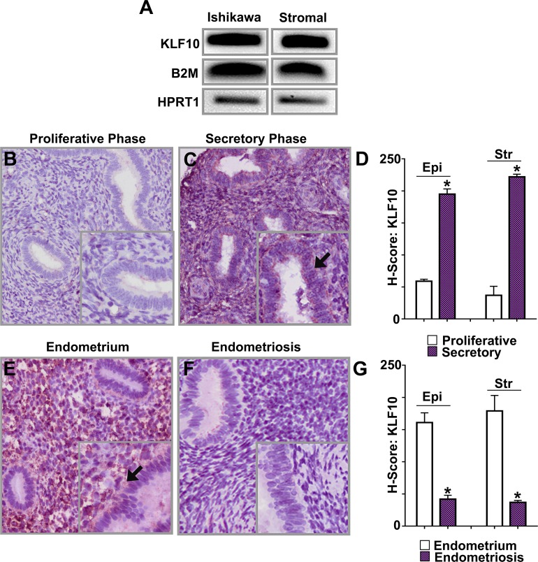
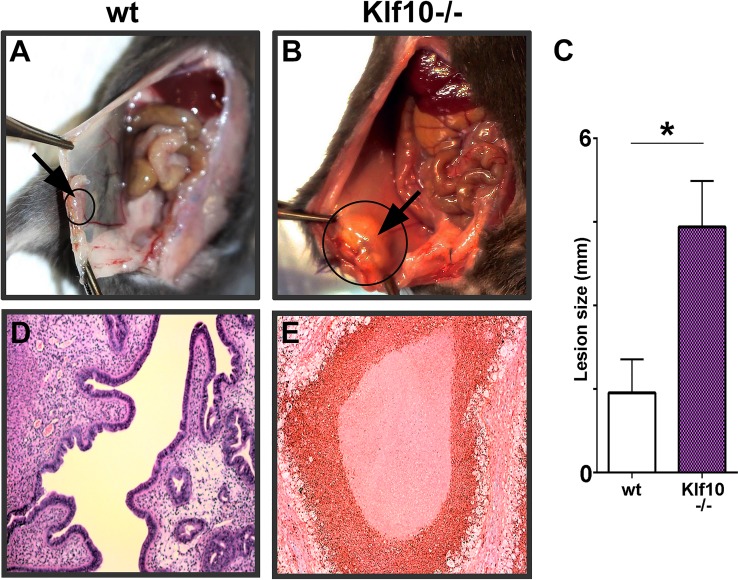
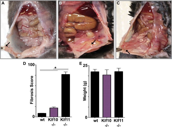
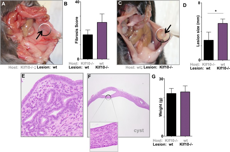
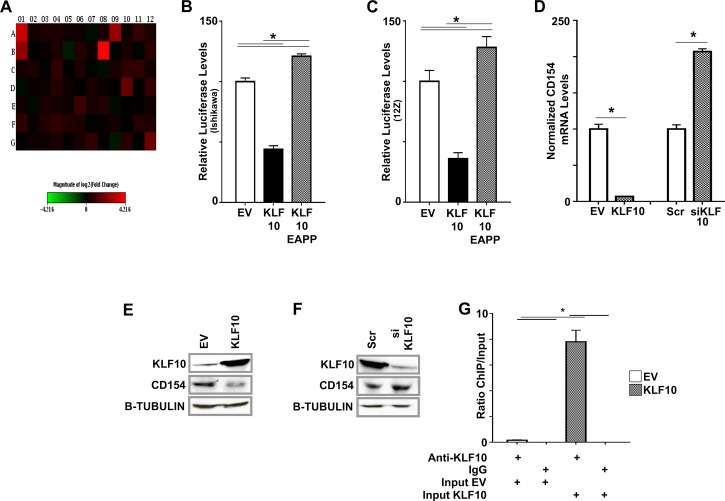
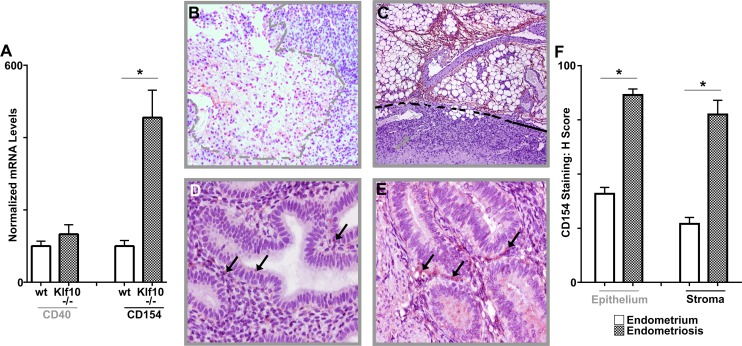
Endometriosis is a highly prevalent, chronic, heterogeneous, fibro-inflammatory disease that remains recalcitrant to conventional therapy. We previously showed that loss of KLF11, a transcription factor implicated in uterine disease, results in progression of endometriosis. Despite extensive homology, co-expression, and human disease association, loss of the paralog Klf10 causes a unique inflammatory, cystic endometriosis phenotype in contrast to fibrotic progression seen with loss of Klf11. We identify here for the first time a novel role for KLF10 in endometriosis. In an animal endometriosis model, unlike wild-type controls, Klf10(-/-) animals developed cystic lesions with massive immune infiltrate and minimal peri-lesional fibrosis. The Klf10(-/-) disease progression phenotype also contrasted with prolific fibrosis and minimal immune cell infiltration seen in Klf11(-/-) animals. We further found that lesion genotype rather than that of the host determined each unique disease progression phenotype. Mechanistically, KLF10 regulated CD40/CD154-mediated immune pathways. Both inflammatory as well as fibrotic phenotypes are the commonest clinical manifestations in chronic fibro-inflammatory diseases such as endometriosis. The complementary, paralogous Klf10 and Klf11 models therefore offer novel insights into the mechanisms of inflammation and fibrosis in a disease-relevant context. Our data suggests that divergence in underlying gene dysregulation critically determines disease-phenotype predominance rather than the conventional paradigm of inflammation being precedent to fibrotic scarring. Heterogeneity in clinical progression and treatment response are thus likely from disparate gene regulation profiles. Characterization of disease phenotype-associated gene dysregulation offers novel approaches for developing targeted, individualized therapy for recurrent and recalcitrant chronic disease.

Keywords: CD154; CD40; CD40 ligand; KLF10; endometriosis; epigenetics; inflammation.


Figures

FIG. 1
FIG. 1
KLF10 expression in epithelial, stromal cells, uterine eutopic endometrium, and endometriosis. A) Expression of KLF10 mRNA was assessed in two cell lines: Ishikawa, a well-differentiated endometrial adenocarcinoma cell line, and human endometrial stromal cells (Stromal). KLF10 was expressed in both cell lines utilizing B2M and HPRT1 as reference controls. To demonstrate KLF10 expression in each of these endometrial cell lines, lysate for RNA or protein extraction was obtained from one 10 cm cell culture dish of confluent cells. B, C) Using a tissue microarray (TMA) consisting of 306 samples of benign uterine endometrium during both the proliferative and secretory phases of the menstrual cycle, immunohistochemistry was performed for KLF10. KLF10 expression was significantly increased in the secretory phase of the menstrual cycle as compared to the proliferative phase (magnification ×200; inset ×400). Inset picture with arrow indicates increased staining. D) H scores were 59 ± 3 and 195 ± 8 in epithelial cells (Epi) and 37 ± 14 and 222 ± 4 in stromal cells (Str) during the proliferative and secretory phases (*P < 0.05 as shown). E, F) KLF10 expression was also evaluated in 28 paired samples of eutopic endometrium and ectopic endometrial implants by immunohistochemistry. Representative samples are shown. KLF10 was expressed in nuclei and cytoplasm of epithelial and stromal cells in both eutopic endometrium as well as in endometriotic implants. KLF10 expression was decreased in endometriotic implants compared to eutopic endometrium (magnification ×200; inset ×400). Inset picture with arrow indicates increased staining. G) Corresponding H scores were: 161 ± 15 and 42 ± 6 in epithelial cells and 179 ± 24 and 37 ± 3 in stromal cells in eutopic endometrium and endometriosis, respectively (*P < 0.05 as shown).
FIG. 2
FIG. 2
Role of KLF10 on disease phenotype and lesion size. A, B, C) Endometriosis was surgically induced in 8-wk-old wt and Klf10−/− female mice (n = 7/group). At induction, 0.5 cm endometrial implants were sutured onto the parietal peritoneum of both groups. Lesion size, morphometry, and disease phenotype were evaluated 3 wk later at necropsy. Peritoneal lesions (circles and arrows) in Klf10−/− animals were larger, cystic, and associated with an inflammatory exudate (B). In contrast, wt mice showed lesion regression and minimal to no fibrosis or inflammatory exudate (A). Average lesion size of Klf10−/− animals was significantly greater (>2 fold) than wt (C). Error bars represent standard deviation (*P < 0.05). D, E) Histochemical analysis using hematoxylin and eosin revealed Klf10−/− lesions to be associated with a large neutrophilic infiltrate and complete obliteration of normal glands and stroma. In contrast, wt lesions had normal appearing glands and stroma. Magnification ×100 (A, B, D, E).
FIG. 3
FIG. 3
Loss of KLF10−/− demonstrates a predominately inflammatory response with minimal fibrosis. Endometriotic lesions in Klf10−/− mice were associated with prolific inflammation with associated infiltrate and minimal associated fibrosis. A) Wild-type animal demonstrating endometrial implant regression (arrow). Lesions were discrete and not physically adherent to surrounding peritoneum or abdominal viscera at time of necropsy. B) Klf10−/− animal demonstrating significant inflammatory infiltrate and minimal fibrosis (arrow: lesion). These lesions were associated with no or minimal easily ruptured adhesions to surrounding structures. There was also no associated mesenteric shortening. C) Klf11−/− animal demonstrating dense fibrosis of bowel to peritoneal lesion (arrow). In contrast to wt and Klf10−/− animals, lesions in Klf11−/− were nondiscrete and encased in adhesions to surrounding peritoneum and abdominal viscera. D) A murine fibrosis adhesion score was utilized to evaluate overall fibrosis in the endometriosis model. Klf11−/− animals demonstrate significantly higher fibrosis scores (>8 fold when compared to wt and >4 fold when compared to Klf10−/−). Error bars represent standard deviation (*P < 0.05). E) Induction of endometriosis, despite varying phenotypes, did not alter animal weights. Error bars represent standard deviation.
FIG. 4
FIG. 4
Progression to endometriosis is lesion rather than host driven. Endometriosis was surgically induced in 8-wk-old wt and Klf10−/− mice (n = 5/group) and transplants were performed by suturing the Klf10−/−implants into wt control animals and vice versa. Lesion size, morphometry, and disease phenotype were evaluated 3 wk later. A) A Klf10−/− animal with wt implants demonstrating lesional regression. B) Klf10−/− animals with wt lesions had a trend toward lower fibrosis scores than wt animals with Klf10−/− lesions. Error bars represent standard deviation. C) Comparatively wt animals with Klf10−/− implants developed larger, cystic lesions. There was, however, no lesional inflammatory exudate as found in the Klf10−/− animals transplanted with Klf10−/−lesions. D) Graph depicts average lesion size for each group. Error bars represent standard deviation (*P < 0.05). E, F) Histochemical analysis of lesions using hematoxylin and eosin stain was performed on both lesion types. Klf10−/− animals with wt-transplanted lesion demonstrated either normal appearing glands and stroma or complete lesional regression (magnification ×200) (E). Wild-type animals with Klf10−/− implants demonstrated large cystic lesions (magnification ×200; inset ×400) (F). G) Animal weights did not differ across experimental groups. Average weight for each group is depicted with error bars representing standard deviation.
FIG. 5
FIG. 5
KLF10 binds to and regulates CD154. A) Heat map showing expression of 84 innate and acquired immune-related genes determined using PCR arrays in Ishikawa cells transfected with Scr and KLF10 siKLF10. The results were normalized to the expression of five housekeeping genes and represent the average fold change from three independent biological replicates. Decrease in KLF10 resulted in a statistically significant elevation in expression of CD40 (A12) and CD154 (B1) at 1.36 and 5.3 fold, respectively (P < 0.05 based on three biological replicates). Complete list of altered genes is provided in Supplemental Table S1. B, C) Ishikawa cells (B) and 12Z cells (C) were cotransfected with pcDNA3/HIS (EV), pcDNA3/HIS-KLF10, or pcDNA3/HIS-KLF10EAPP and a pGL4/CD154-promoter-reporter construct. KLF10 repressed CD154-promoter luciferase activity compared to EV (*P < 0.05 compared to EV). In contrast, KLF10EAPP derepressed and thus activated CD154 promoter luciferase expression (*P < 0.05, compared to KLF10 and EV as indicated). Luciferase levels were normalized to total lysate protein concentration. Assays were repeated in triplicate three times. D) Overexpression of KLF10 in Ishikawa cells decreased normalized CD154 mRNA expression 9 fold compared to EV. In contrast, CD154 mRNA expression levels were significantly increased in Ishikawa cells transfected with KLF10 siRNA compared to Scr control. CD154 mRNA expression was normalized to five housekeeping genes (mean expression levels ± SEM shown, *P < 0.05). E) KLF10 overexpression in Ishikawa cells transfected with pcDNA3/HIS-KLF10 cognately suppressed CD154 protein expression compared to corresponding EV. Beta-TUBULIN was used as a loading control. F) Conversely, suppressed transcription factor expression in cells transfected with KLF10siRNA was associated with increased CD154 protein expression compared to that in cells transfected with scrambled control (Scr). Beta-TUBULIN was used as a loading control. G) Chromatin immunoprecipitation (ChIP) assay was used to determine direct KLF10 binding to the region −600 to −401 of the CD154 promoter in Ishikawa cells. Promoter binding was detected by anti-KLF10 but not a control species- and isotype-specific IgG. Promoter-transcription factor binding was increased nearly 8 fold in cells transfected with pcDNA3/His KLF10 compared to EV. Levels were normalized to input (diluted 1:100). *P < 0.05 for comparisons of normalized binding levels in EV or KLF10-transfected cells analyzed by ChIP using anti-KLF10 or IgG as well as for comparison of normalized binding levels in EV and KLF10-transfected cells analyzed by ChIP using anti-KLF10 as indicated.
FIG. 6
FIG. 6
Role of CD154 in murine endometriotic implants and human endometriosis. A) CD154 mRNA expression levels were determined from one of two endometriotic implants in each animal (Klf10−/− and wt, n = 7). CD154 expression levels were increased >4 fold in implants from Klf10−/− animals compared to wt (*P < 0.05; error bars represent SEM). B, C) CD154 expression in wt (B) and Klf10−/− (C) implants demonstrate increased expression in Klf10−/− lesions particularly in vascular areas surrounding the neutrophilic infiltrate. Dashed lines indicate lesion extent and region of neutrophilic infiltrate. Magnification ×100. D, E) CD154 expression was also evaluated by immunohistochemistry in a tissue microarray (TMA) of 28 patients comparing eutopic (D) and ectopic endometria (E). CD154 expression was higher in ectopic compared to corresponding eutopic endometrium (arrows). Magnification ×400. F) Differences in CD154 expression were reflected in their H scores of 81 ± 6 and 163 ± 5.5 for epithelial CD154 expression in eutopic and ectopic endometrium, respectively, and 54 ± 6 and 155 ± 13 for stromal CD154 expression in eutopic and ectopic endometrium, respectively (*P < 0.05).

Similar articles

Cited by

References

    1. Giudice LC, Kao LC. Endometriosis. Lancet. 2004;364:1789–1799. - PubMed
    1. Bonnefond A, Lomberk G, Buttar N, Busiah K, Vaillant E, Lobbens S, Yengo L, Dechaume A, Mignot B, Simon A, Scharfmann R, Neve B, et al. Disruption of a novel Kruppel-like transcription factor p300-regulated pathway for insulin biosynthesis revealed by studies of the c.-331 INS mutation found in neonatal diabetes mellitus. J Biol Chem. 2011;286:28414–28424. - PMC - PubMed
    1. Daftary GS, Zheng Y, Tabbaa ZM, Schoolmeester JK, Gada RP, Grzenda AL, Mathison AJ, Keeney GL, Lomberk GA, Urrutia R. A novel role of the Sp/KLF transcription factor KLF11 in arresting progression of endometriosis. Plos One. 2013;8:e60165. - PMC - PubMed
    1. Mathison A, Grzenda A, Lomberk G, Velez G, Buttar N, Tietz P, Hendrickson H, Liebl A, Xiong YY, Gores G, Fernandez-Zapico M, Larusso NF, et al. Role for Krüppel-like transcription factor 11 in mesenchymal cell function and fibrosis. PLoS One. 2013;8:e75311. - PMC - PubMed
    1. Yin P, Lin Z, Reierstad S, Wu J, Ishikawa H, Marsh EE, Innes J, Cheng Y, Pearson K, Coon JS V, Kim JJ, Chakravarti D, Bulun SE. Transcription factor KLF11 integrates progesterone receptor signaling and proliferation in uterine leiomyoma cells. Cancer Res. 2010;70:1722–1730. - PMC - PubMed

Publication types

MeSH terms