. 2017 Jan;47(1):22-32.
doi: 10.1097/SHK.0000000000000713.

Retinoic Acid Improves Incidence and Severity of Necrotizing Enterocolitis by Lymphocyte Balance Restitution and Repopulation of LGR5+ Intestinal Stem Cells

Affiliations

Retinoic Acid Improves Incidence and Severity of Necrotizing Enterocolitis by Lymphocyte Balance Restitution and Repopulation of LGR5+ Intestinal Stem Cells

Diego F Niño et al. Shock. 2017 Jan.

Abstract

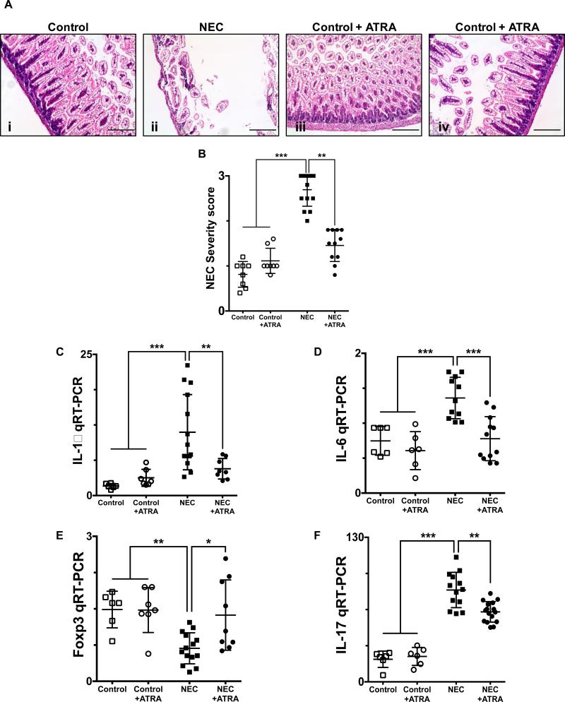
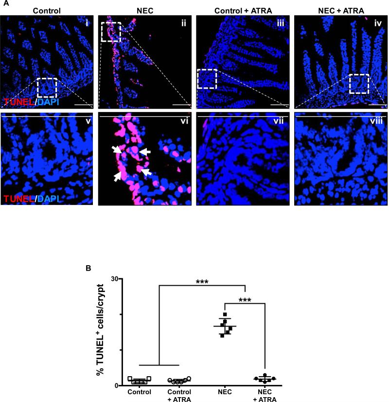
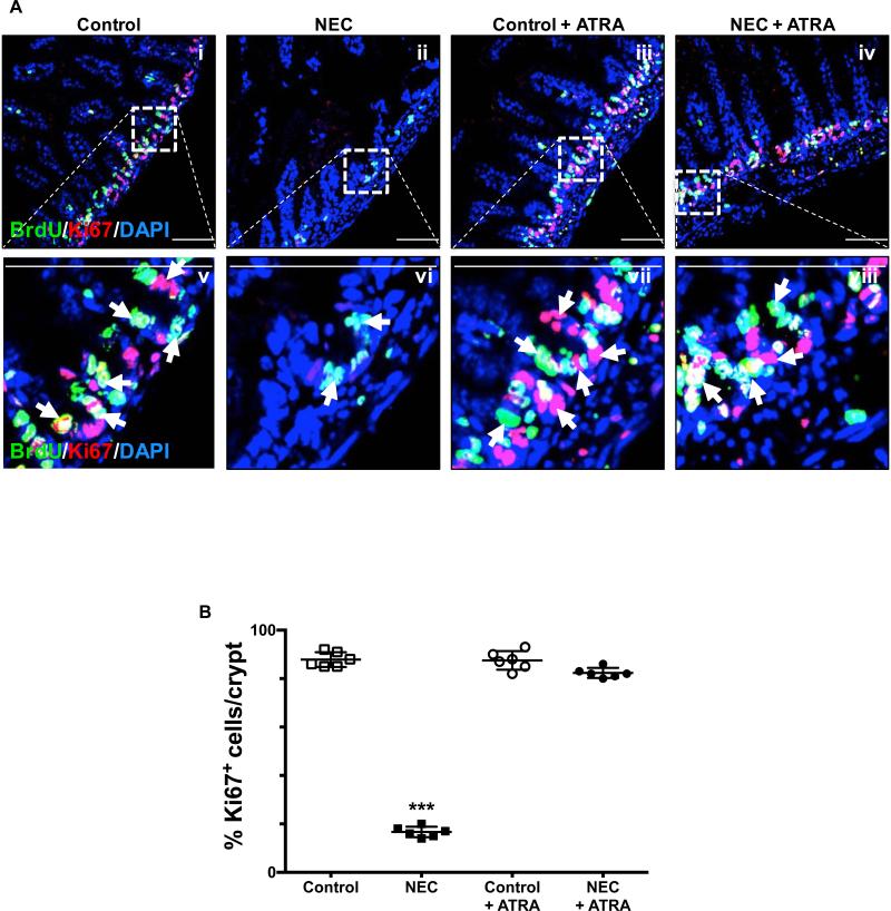
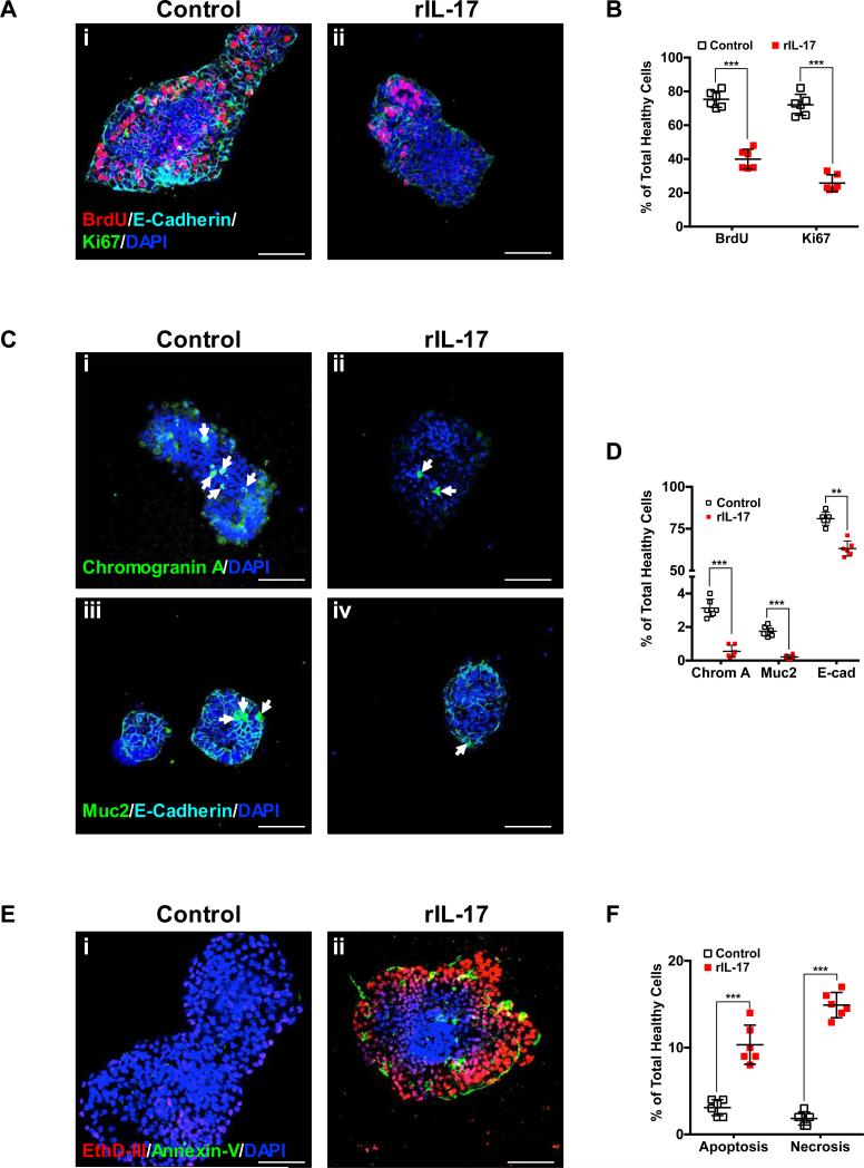
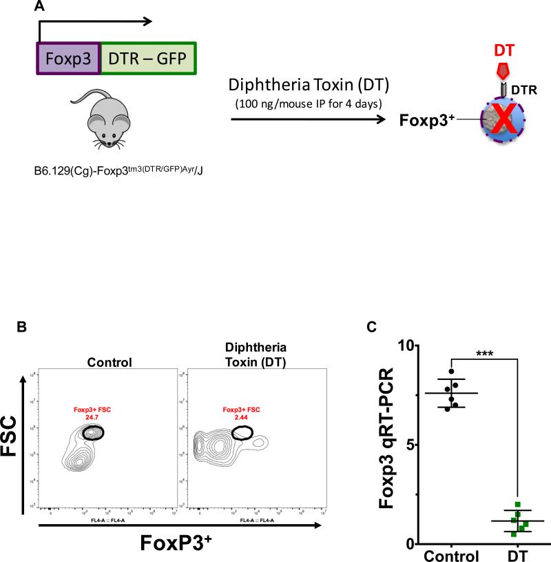
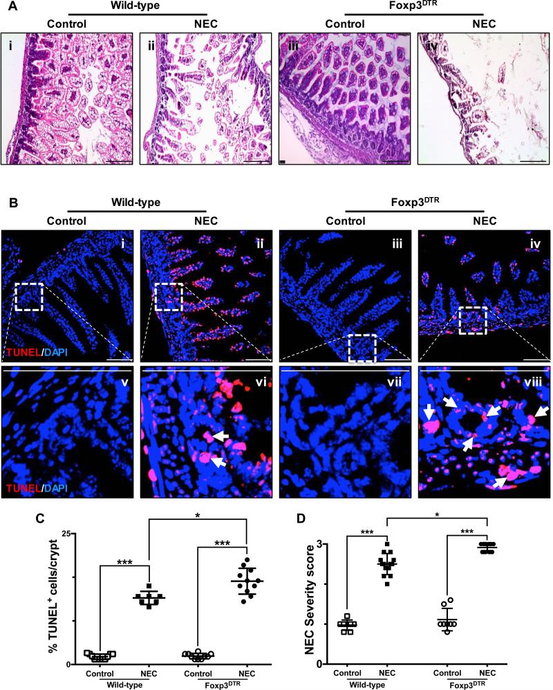
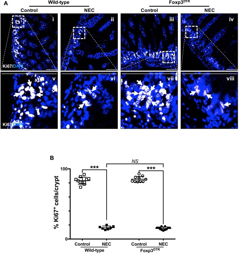
Necrotizing enterocolitis (NEC) is the most devastating gastrointestinal disease of the premature infant. We have recently shown that NEC development occurs after an increase in proinflammatory CD4Th17 (Th17) cells and reduced anti-inflammatory forkhead box P3 regulatory T cells (Tregs) to the premature small intestine of mice and humans, which can be experimentally reversed in mice by administration of all-trans retinoic acid (ATRA). We have also shown that NEC is characterized by apoptosis of Lgr5-positive intestinal stem cells (ISCs-Lgr5 cells) within the crypts of Lieberkühn, which are subsequently essential for intestinal homeostasis. We now hypothesize that the normal lymphocyte balance within the lamina propria of the intestine can be achieved via administration of ATRA which restores mucosal integrity by preventing the loss of ISCs. Using both in vivo and in vitro strategies, we now demonstrate that Th17 recruitment and Treg depletion lead to increased apoptosis within ISC niches, significantly impairing proliferative capacity and mucosal healing. ATRA exerted its protective effects by preventing T cell imbalance, ultimately leading to the protection of the ISC pool preventing the development of NEC in mice. These findings raise the exciting possibility that dietary manipulations could prevent and treat NEC by modulating lymphocyte balance and the ISC pool within the newborn small intestine.


Figures

Fig. 1
Fig. 1. Enteral administration of retinoic acid prevents the development of necrotizing enterocolitis
A, H&E sections of the terminal ileum obtained from control mice that did not (i) or did (iii) receive ATRA (50 μg/mouse orally per day) and mice submitted to experimental NEC in the absence (ii) or presence (iv) of ATRA. Histological evaluation demonstrates a preserved intestinal mucosa in mice that received ATRA during the course of induction of experimental NEC. B, Injury severity score as determined by a pathologist blinded to the experimental conditions, demonstrates a significant decrease in the incidence and severity of experimental NEC in the presence of ATRA intervention. Gene expression analysis of the terminal ileum (C – D) or lamina propria isolated CD4+ T cells (E – F), obtained from control mice and mice submitted to experimental NEC in the absence or presence of ATRA. Inflammatory mediators IL-1β (C) and IL-6 (D) demonstrate a significant decrease in the inflammatory response associated with NEC in animals that received ATRA. Furthermore, CD4+ T cell gene expression profile was evaluated using Foxp3 (E) and IL-17 (F) expression; demonstrating preserved levels of Tregs (E) and absence of Th17 induction (F) among animals that were exposed to NEC and received oral ATRA. Scale bar = 50μm. Data depicted is presented as mean ± SD and represents 3 independent experiments with at least 10 mice per group. *P ≤ 0.05, **P ≤ 0.001, ***P ≤ 0.0001 determined by ANOVA followed by Tukey's multiple comparisons test.
Fig. 2
Fig. 2. Enteral administration of retinoic acid attenuates the effect of necrotizing enterocolitis by preventing the induction of apoptosis of crypt-based intestinal stem cells
A, confocal images of TUNEL stained sections of the terminal ileum obtained from control mice that did not (i and v) or did (iii and vii) receive ATRA (50 μg/mouse orally per day) and mice submitted to experimental NEC in the absence (ii and vi) or presence (iv and viii) of ATRA. The outlined areas in i – iv are shown at a higher magnification in v – viii. Apoptotic cells within the crypts are identified by arrows and demonstrate the significant effect that ATRA has on the induction of NEC by preventing cell death within the ISC population. B, TUNEL staining intensity was quantified as described in the methods section and expressed as a percentage of the total number of cells identified within the crypts per high-magnification field. Scale bar = 50μm. Data depicted is presented as mean ± SD and represents 3 independent experiments with at least 10 mice per group. ***P ≤ 0.0001 determined by ANOVA followed by Tukey's multiple comparisons.
Fig. 3
Fig. 3. Enteral administration of retinoic acid preserves the proliferative capacity of crypt-based intestinal stem cells
A, the proliferative capacity of crypt-based ISCs was assessed by BrdU label incorporation and Ki67 staining of terminal ileum sections obtained from control mice that did not (i and v) or did (iii and vii) receive ATRA (50 μg/mouse orally per day) and mice submitted to experimental NEC in the absence (ii and vi) or presence (iv and viii) of ATRA. The outlined areas in i – iv are shown at a higher magnification in v – viii. Proliferating cells are indicated by arrows and demonstrate a conserved stable pool of dividing cells within the crypts. B, Ki67 staining was quantified as described in the methods section and is expressed as a percentage of the total number of cells identified within the crypts per high-magnification field. Scale bar = 50μm. Data depicted is presented as mean ± SD and represents 3 independent experiments with at least 10 mice per group. ***P ≤ 0.0001 determined by ANOVA followed by Tukey's multiple comparisons.
Fig. 4
Fig. 4. IL-17 is detrimental to ISC homeostasis by inhibiting cell proliferation and differentiation and leading to apoptosis/necrosis
The effects of the main cytokine released by Th17 cells (i.e. IL-17) on ISC biology was evaluated using enteroids generated from mouse ileum ISCs and exposed to recombinant IL-17 (rIL-17, 100 ng/mL for 6 h). Cell proliferation (A and B), cell differentiation (C and D) and apoptosis/necrosis (E and F) were assessed as described in the methods section. A, cell proliferation was evaluated in control and rIL-17 exposed enteroids using incorporation of BrdU label as well as expression of the proliferation marker Ki67. Representative confocal microscopy images of control (i) and rIL-17 treated (ii) enteroids are shown. C, cell differentiation was assessed by immunostaining for the enteroendocrine marker chromogranin A, the goblet cell marker muc-2 and the enterocyte marker E-cadherin. Representative confocal microscopy images of control (i and iii) and rIL-17 treated (ii and iv) enteroids are shown. Chromogranin A (i and ii) and muc-2 (iii and iv) positive cells are indicated by arrows. E, apoptosis and necrosis were assessed using the apoptotic cell marker Annexin V and necrotic cells were identified by their permeability to the nucleic acid probe ethidium homodimer III (EthD-III). Representative confocal microscopy images of control (i) and rIL-17 treated (ii) enteroids are shown. B, D and F, quantification of fluorescence intensity and cell number was performed using FIJI software and depicted as the average percentage of the total number of healthy cells. The overall effect of IL-17 on enteroids derived from ISCs is characterized by a significant decrease in cellular proliferation and differentiation as well as increased apoptosis/necrosis. All images depicted are representative of three separate experiments with at least five enteroids analyzed/group. Scale bar = 100μm. Data depicted is presented as mean ± SD. **P ≤ 0.01, ***P ≤ 0.001 determined by Student's t test.
Fig. 5
Fig. 5. Diphtheria toxin-induced depletion of Foxp3+ regulatory T cells
A, schematic diagram depicts the experimental strategy as described in the methods section utilized for the depletion of Foxp3+ regulatory T cells (Tregs) in Foxp3DTR mice. Depletion of this cell population was confirmed by flow cytometry (B) and qRT-PCR (C). B, representative plot demonstrates the depletion of Tregs within the lamina propria of the terminal ileum obtained from Foxp3DTR mice that received diphtheria toxin (DT 100 ng/mouse IP, daily for 4 days). Flow cytometric analysis was performed on CD4+ T cells, which were magnetically isolated by positive selection using anti-CD4 beads. Control samples were obtained from mice that received saline. C, depletion of Tregs was further confirmed by qRT-PCR analysis of Foxp3 expression in the purified cell population obtained after magnetic isolation as described above. Data depicted in (C) is presented as mean ± SD and represents 3 independent experiments with at least 6 mice per group. ***P ≤ 0.0001 determined by Student's t test.
Fig. 6
Fig. 6. Depletion of regulatory T cells exacerbates the development of experimental necrotizing enterocolitis
A, H&E sections of the terminal ileum obtained from wild-type C57Bl/6 and Foxp3+DTR mice submitted to experimental NEC (ii and iv, respectively) or breast-fed controls (i and iii, respectively). B, confocal images of TUNEL stained sections of the terminal ileum obtained from wild-type C57Bl/6 and Foxp3+DTR mice submitted to experimental NEC (ii, vi and iv, viii respectively) or breast-fed controls (i, v and iii, vii respectively). The outlined areas in i – iv are shown at a higher magnification in v – viii. Apoptotic cells within the crypts are identified by arrows and demonstrate the significant effect that depletion of Foxp3+ T cells has on the induction and severity of NEC, exacerbating the damage to the crypt-based ISC population. TUNEL staining intensity (C) was quantified as described in the methods section and is expressed as a percentage of the total number of cells identified within the crypts per high-magnification field. D, injury severity score as determined by a pathologist blinded to the experimental conditions, demonstrates a significant increase in the incidence and severity of experimental NEC among Foxp3+DTR mice. Scale bar = 50μm. Data depicted is presented as mean ± SD and represents 3 independent experiments with at least 10 mice per group. *P ≤ 0.05, ***P ≤ 0.0001 determined by ANOVA followed by Tukey's multiple comparisons.
Fig. 7
Fig. 7. Depletion of regulatory T cells exacerbates the development of experimental necrotizing enterocolitis by decreasing the proliferative capacity of crypt-based intestinal stem cells
A, the proliferative capacity of crypt-based ISCs was assessed by Ki67 staining of terminal ileum sections obtained from wild-type C57Bl/6 and Foxp3+DTR mice submitted to experimental NEC (ii, vi and iv, viii respectively) or breast-fed controls (i, v and iii, vii respectively). The outlined areas in i – iv are shown at a higher magnification in v – viii. Proliferating cells are indicated by arrows and demonstrate a significant decrease of dividing cells within the crypts of mice submitted to NEC. B, Ki67 staining was quantified as described in the methods section and expressed as a percentage of the total number of cells identified within the crypts per high-magnification field. Scale bar = 50μm. Data depicted is presented as mean ± SD and represents 3 independent experiments with at least 10 mice per group. ***P ≤ 0.0001 determined by ANOVA followed by Tukey's multiple comparisons.

Similar articles

Cited by

References

    1. Nino DF, Sodhi CP, Hackam DJ. Necrotizing enterocolitis: mechanisms and management. Nat Rev Gastroenterol Hepatol. 2016 In Press. - PMC - PubMed
    1. Neu J, Walker WA. Necrotizing enterocolitis. N Engl J Med. 2011;364(3):255–264. - PMC - PubMed
    1. Stoll BJ, Hansen NI, Bell EF, Walsh MC, Carlo WA, Shankaran S, Laptook AR, Sanchez PJ, Van Meurs KP, Wyckoff M, et al. Trends in Care Practices, Morbidity, and Mortality of Extremely Preterm Neonates, 1993-2012. JAMA. 2015;314(10):1039–1051. - PMC - PubMed
    1. Stey A, Barnert ES, Tseng CH, Keeler E, Needleman J, Leng M, Kelley-Quon LI, Shew SB. Outcomes and costs of surgical treatments of necrotizing enterocolitis. Pediatrics. 2015;135(5):e1190–1197. - PMC - PubMed
    1. Anand RJ, Leaphart CL, Mollen KP, Hackam DJ. The role of the intestinal barrier in the pathogenesis of necrotizing enterocolitis. Shock. 2007;27(2):124–133. - PubMed

Publication types