. 2016 Nov;30(11):3759-3770.
doi: 10.1096/fj.201600353R. Epub 2016 Aug 3.

The circadian clock regulates inflammatory arthritis

Affiliations

The circadian clock regulates inflammatory arthritis

Laura E Hand et al. FASEB J. 2016 Nov.

Abstract

There is strong diurnal variation in the symptoms and severity of chronic inflammatory diseases, such as rheumatoid arthritis. In addition, disruption of the circadian clock is an aggravating factor associated with a range of human inflammatory diseases. To investigate mechanistic links between the biological clock and pathways underlying inflammatory arthritis, mice were administered collagen (or saline as a control) to induce arthritis. The treatment provoked an inflammatory response within the limbs, which showed robust daily variation in paw swelling and inflammatory cytokine expression. Inflammatory markers were significantly repressed during the dark phase. Further work demonstrated an active molecular clock within the inflamed limbs and highlighted the resident inflammatory cells, fibroblast-like synoviocytes (FLSs), as a potential source of the rhythmic inflammatory signal. Exposure of mice to constant light disrupted the clock in peripheral tissues, causing loss of the nighttime repression of local inflammation. Finally, the results show that the core clock proteins cryptochrome (CRY) 1 and 2 repressed inflammation within the FLSs, and provide novel evidence that a CRY activator has anti-inflammatory properties in human cells. We conclude that under chronic inflammatory conditions, the clock actively represses inflammatory pathways during the dark phase. This interaction has exciting potential as a therapeutic avenue for treatment of inflammatory disease.-Hand, L. E., Hopwood, T. W., Dickson, S. H., Walker, A. L., Loudon, A. S. I., Ray, D. W., Bechtold, D. A., Gibbs, J. E. The circadian clock regulates inflammatory arthritis.

Keywords: cryptochrome; diurnal; fibroblast-like synoviocyte; rheumatoid arthritis.


Figures

Figure 1.
Figure 1.
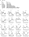
Diurnal variation in the disease pathology of CIA. A) Disease incidence, paw swelling, and disease severity scored by DPI in the murine CIA model (n = 19). B) Paw swelling of inflamed limbs (scoring 3 or 4) was measured in a subset of animals (n = 3 animals) at 8 consecutive times 12 h apart. For each inflamed paw, measurements were normalized to the first measurement at ZT6 (100%) (repeated-measures Student’s t test). C) Serum obtained from serial tail bleeds at ZT6 and -18 on DPI 30 from CIA (n = 7–8), vehicle (n = 4), or naive (n = 3–4) mice were analyzed for cytokine levels, 2-way ANOVA with post hoc Tukey. *P ≤ 0.05; ***P ≤ 0.005.
Figure 2.
Figure 2.
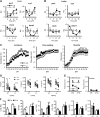
Expression of inflammatory markers and clock genes in inflamed limbs. A) Relative quantification of inflammatory gene transcripts in limbs harvested from naive (n = 4) or CIA (n = 5) animals at ZT0 (normalized to expression in naïve limbs; Student’s t test). B) Cytokine transcript levels were measured in limbs harvested from CIA mice at 4 time points across the day (n = 5). Values are made relative to samples collected from naive mice at ZT0. Kruskal-Wallis and post hoc Dunn comparing CIA values to naive values at ZT0. C) Clock gene transcript levels were measured in limbs harvested from CIA mice at 4 time points across the day (n = 5). Values are made relative to samples collected at ZT0. One-way ANOVA and post hoc Tukey. aSignificant change vs. ZT6. bSignificant change vs. ZT12. *P ≤ 0.05; **P ≤ 0.01.
Figure 3.
Figure 3.
Circadian disruption abrogates rhythmic inflammation in the limbs. In these studies, samples were collected from LL and LD mice at the same time, with ZT in LD mice used as temporal markers. A, B) Control experiments confirmed that maintenance of animals in LL flattened circadian rhythms within peripheral organs (A, liver) and within the limbs (B), n = 3–4/time point. C) Housing mice in LL before and during CIA induction had no effect on the incidence, paw swelling or severity of CIA, n = 29/group. D) Serial blood samples taken from arthritic animals (n = 9/group) at 2 opposing time points 12 h apart (ZT6 and -18) were analyzed for cytokine levels with a BioPlex assay; paired samples are joined by lines (paired Student’s t test). E) In a separate cohort of animals, paw size was measured in severely inflamed paws (score 3–4, n = 4–5 animals) at 38 DPI (and in uninflamed paws as controls; n = 5–7 animals) at 2 time points 12 h apart. F) Cytokine transcript levels were analyzed in limbs harvested at ZT6 or -18 from CIA mice housed in LD or LL (n = 4–8) conditions; values were normalized to samples collected at ZT6 from nonarthritic mice housed in LD (2-way ANOVA and post hoc Tukey). *P ≤ 0.05, **P ≤ 0.01 for time-of-day differences (ZT6 vs. -18) within treatment groups. +P ≤ 0.05, ++P ≤ 0.01 for significant lighting effect (LD vs. LL) at the same time point.
Figure 4.
Figure 4.
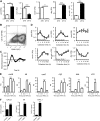
FLSs as rhythmic effector cells in the joints. A) FLSs sorted from naïve limbs of mice euthanized at ZT0 (n = 4) or ZT12 (n = 5) were analyzed for expression of clock genes. Clock gene expression was normalized to levels at ZT0, comparisons were made by using the Student’s t test. B) Flow cytometric analysis of expression of CD90.2 (FLS marker) and CD11b (macrophage marker) in synovial fibroblasts cultured from DBA/1 mice indicating population purity. C) FLSs isolated from PER2::luc mice showed rhythmic bioluminescent output under PMTs with a mean period of 24.8 ± 0.1 h (n = 6). D) FLSs cultured from DBA/1 mice (naive) showed circadian rhythmicity in core clock genes after synchronization (n = 3). E) Murine (DBA/1) synovial fibroblasts were challenged with TNFα for 30 min to 2 h and expression of inflammatory cytokines quantified relative to naive controls (n = 3); ifnγ and ifnβ levels were undetectable (1-way ANOVA and post hoc Bonferroni test). F) Murine (DBA/1) synovial fibroblasts were challenged with TNFα for 24 h. Cell supernatants were analyzed for cytokine production, Student’s t tests (n = 3); IL1β and IL10 levels were undetectable. *P ≤ 0.05; **P ≤ 0.01; ***P ≤ 0.005.
Figure 5.
Figure 5.
CRY represses inflammation. FLSs derived from CRY DKO mice or WT counterparts were stimulated with TNFα (10 ng/ml). A) Proinflammatory cytokine gene transcripts were quantified after 2 h relative to expression in naïve WT cells (2-way ANOVA and post hoc Bonferroni test). B) Protein levels in cell supernatants were quantified by ELISA after TNFα stimulation for 6 h, 2-way ANOVA, post hoc Bonferroni. C, D) FLSs derived from DBA/1 mice were transfected with siRNAs targeting cry1 and cry2, or both, and stimulated with TNFα; qPCR was performed to confirm siRNA knockdown of target genes (C) and quantify proinflammatory cytokine gene transcripts (D). Data are relative to expression in control untreated cells, representative of 3 independent trials (1-way ANOVA and post hoc Bonferroni test). **P ≤ 0.01; ***P ≤ 0.005.
Figure 6.
Figure 6.
Anti-inflammatory properties of CRY activation on murine and human FLSs. A) Human FLSs were synchronized and then harvested every 4 h to assess rhythmic expression of core clock genes (n = 3). Human FLSs were stimulated with TNFα for 2 h with or without pretreatment for 1 h with 8 μM KL001. qPCR was performed to quantify clock gene expression (B) and proinflammatory cytokine gene transcripts (C). Data are relative to expression in control-treated cells, representative of 3 independent trials (Student’s t test). D) FLSs cells (passage 3) derived from DBA/1 mice were stimulated with TNFα for 2 h with or without pretreatment for 1 h with 8 μM KL001. qPCR was performed to quantify proinflammatory cytokine gene transcripts. Data are a percentage of cytokine transcription after TNFα stimulation to account for experimental variability between repeats (Student’s t test). E) CXCL1, IL6, and CXCL5 protein production by FLSs in response to TNFα stimulation for 4 h in the absence and presence of KL001 were quantified by Bioplex assay (n = 3); IL1β protein was not detectable (1-way ANOVA and post hoc Bonferroni test). *P ≤ 0.05; **P ≤ 0.01; ***P ≤ 0.005 vs. the control unstimulated group.

References

    1. Hastings M. H., Maywood E. S., Reddy A. B. (2008) Two decades of circadian time. J. Neuroendocrinol. 20, 812–819 - PubMed
    1. Labrecque N., Cermakian N. (2015) Circadian clocks in the immune system. J. Biol. Rhythms 30, 277–290 - PubMed
    1. Gibbs J. E., Blaikley J., Beesley S., Matthews L., Simpson K. D., Boyce S. H., Farrow S. N., Else K. J., Singh D., Ray D. W., Loudon A. S. (2012) The nuclear receptor REV-ERBα mediates circadian regulation of innate immunity through selective regulation of inflammatory cytokines. Proc. Natl. Acad. Sci. USA 109, 582–587 - PMC - PubMed
    1. Keller M., Mazuch J., Abraham U., Eom G. D., Herzog E. D., Volk H. D., Kramer A., Maier B. (2009) A circadian clock in macrophages controls inflammatory immune responses. Proc. Natl. Acad. Sci. USA 106, 21407–21412 - PMC - PubMed
    1. Arjona A., Sarkar D. K. (2005) Circadian oscillations of clock genes, cytolytic factors, and cytokines in rat NK cells. J. Immunol. 174, 7618–7624 - PubMed

Publication types

LinkOut - more resources