. 2016 Aug 4;15(1):397.
doi: 10.1186/s12936-016-1449-5.

Anti-erythrocyte antibodies may contribute to anaemia in Plasmodium vivax malaria by decreasing red blood cell deformability and increasing erythrophagocytosis

Affiliations

Anti-erythrocyte antibodies may contribute to anaemia in Plasmodium vivax malaria by decreasing red blood cell deformability and increasing erythrophagocytosis

Luiza Carvalho Mourão et al. Malar J. .

Abstract

Background: Plasmodium vivax accounts for the majority of human malaria infections outside Africa and is being increasingly associated in fatal outcomes with anaemia as one of the major complications. One of the causes of malarial anaemia is the augmented removal of circulating non-infected red blood cells (nRBCs), an issue not yet fully understood. High levels of auto-antibodies against RBCs have been associated with severe anaemia and reduced survival of nRBCs in patients with falciparum malaria. Since there are no substantial data about the role of those antibodies in vivax malaria, this study was designed to determine whether or not auto-antibodies against erythrocytes are involved in nRBC clearance. Moreover, the possible immune mechanisms elicited by them that may be associated to induce anaemia in P. vivax infection was investigated.

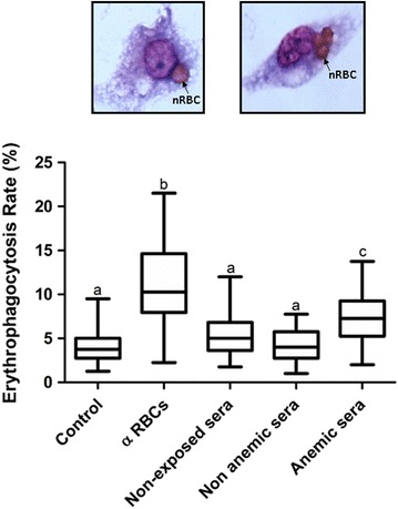
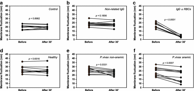
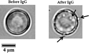
Methods: Concentrations of total IgG were determined by sandwich ELISA in sera from clinically well-defined groups of P. vivax-infected patients with or without anaemia and in healthy controls never exposed to malaria, whereas the levels of specific IgG to nRBCs were determined by cell-ELISA. Erythrophagocytosis assay was used to investigate the ability of IgGs purified from each studied pooled sera in enhancing nRBC in vitro clearance by THP-1 macrophages. Defocusing microscopy was employed to measure the biomechanical modifications of individual nRBCs opsonized by IgGs purified from each group.

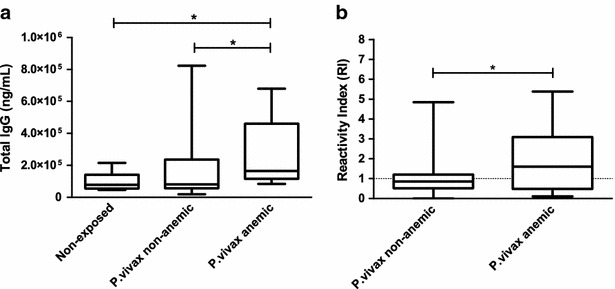
Results: Anaemic patients had higher levels of total and specific anti-RBC antibodies in comparison to the non-anaemic ones. Opsonization with purified IgG from anaemic patients significantly enhanced RBCs in vitro phagocytosis by THP-1 macrophages. Auto-antibodies purified from anaemic patients decreased the nRBC dynamic membrane fluctuations suggesting a possible participation of such antibodies in the perturbation of erythrocyte flexibility and morphology integrity maintenance.

Conclusions: These findings revealed that vivax-infected patients with anaemia have increased levels of IgG auto-antibodies against nRBCs and that their deposition on the surface of non-infected erythrocytes decreases their deformability, which, in turn, may enhance nRBC clearance by phagocytes, contributing to the anaemic outcome. These data provide insights into the immune mechanisms associated with vivax malaria anaemia and may be important to the development of new therapy and vaccine strategies.

Keywords: Anaemia; Auto-antibodies; Defocusing microscopy; Erythrophagocytosis; Non-infected RBC; Plasmodium vivax.


Figures

Fig. 1
Fig. 1
Associations between antibody responses and clinical status. a The concentrations of total IgG and b the levels of IgG against surface molecules of non-infected red blood cells (nRBCs) were evaluated in sera from healthy individuals (n = 11) and in sera from non-anaemic (n = 119) or anaemic P. vivax-infected patients (n = 11) by sandwich and cell-ELISA, respectively. The results are shown as median values and interquartile ranges. p values were determined using nonparametric tests: a Kruskal–Wallis followed by a post hoc Dunn’s multiple comparison test or b Mann–Whitney test. Asterisks indicate statistically significant difference (p value <0.05). Reactivity index (RI) was calculated as the ratio between the mean OD generated by each duplicate and the mean OD plus three standard deviations of samples from 11 malaria-naïve blood donors never exposed to malaria
Fig. 2
Fig. 2
Evaluation of the phagocytic uptake, by THP-1 cells, of non-infected RBCs after their opsonization with different antibodies. nRBCs were isolated from a O+ healthy volunteer and after washing, they were opsonized with different antibodies: IgG against human red blood cells (α RBCs), IgG purified from healthy donors, IgG purified from non-anaemic patients with vivax malaria or IgG purified from patients with vivax malaria and anaemia. A group of nRBCs incubated with PBS was included for control. The combined result of six independent experiments is shown. Erythophagocytosis rate was calculated by counting the number of ingested nRBC on 400 phagocytic cells, under oil immersion light microscopy at 1000× magnification. Data are shown as box-and-whiskers plots, representing interquartile and complete ranges, with the horizontal line in each box indicating the median. p values were determined using ANOVA with Tukey’s post hoc test, in which all groups were pair-wised for statistical significance. Alphabetic letters above each box plot represent the results of Tukey’s multiple comparison test. Erythrophagocytosis rates that are not significantly different one from each other are represented with the same letter whereas unique (unshared) alphabetic letter indicate a significant different rate (p value <0.01). Top two representative light micrographs of ingested nRBCs within THP-1 activated monocytic cells
Fig. 3
Fig. 3
Effects of antibodies on the dynamics fluctuations of nRBC membrane. Modifications in the amplitude of membrane fluctuations of single human RBCs from a healthy donor were examined by DM before and 30 min after the addition of different antibodies: a control, b anti-bacteriophage monoclonal antibody produced in rabbit (non-related IgG), c anti-red blood cell antibody, d IgG purified from sera of healthy individuals, e IgG purified from sera of non-anaemic patients with vivax malaria, and f IgG purified from sera of subjects infected by P. vivax and with anaemia. For each assayed antibody, ten RBCs were evaluated. p values were determined using a paired t test
Fig. 4
Fig. 4
RBCs produce microparticles (indicated by arrows) after the addition of anti-red blood cell antibody (2 % v/v) in the cell suspension. The images were obtained using DM. A single human nRBC from a healthy donor was examined before and 30 min after the addition of the anti-red blood cell antibody

Similar articles

Cited by

References

    1. Genton B, D’Acremont V, Rare L, Baea K, Reeder JC, Alpers MP, et al. Plasmodium vivax and mixed infections are associated with severe malaria in children: a prospective cohort study from Papua New Guinea. PLoS Med. 2008;5:e127. doi: 10.1371/journal.pmed.0050127. - DOI - PMC - PubMed
    1. Tjitra E, Anstey NM, Sugiarto P, Warikar N, Kenangalem E, Karyana M, et al. Multidrug-resistant Plasmodium vivax associated with severe and fatal malaria: a prospective study in Papua. Indonesia PLoS Med. 2008;5:e128. doi: 10.1371/journal.pmed.0050128. - DOI - PMC - PubMed
    1. Alexandre MA, Ferreira CO, Siqueira AM, Magalhães BL, Mourão MPG, Lacerda MV, et al. Severe Plasmodium vivax malaria Brazilian Amazon. Emerg Infect Dis. 2010;16:1611–1614. doi: 10.3201/eid1610.100685. - DOI - PMC - PubMed
    1. Wickramasinghe SN, Abdalla SH. Blood and bone marrow changes in malaria. Baillieres Best Pract Res Clin Haematol. 2000;13:277–299. doi: 10.1053/beha.1999.0072. - DOI - PubMed
    1. Van Hensbroek MB, Calis JCJ, Phiri KS, Vet R, Munthali F, Kraaijenhagen R, et al. Pathophysiological mechanisms of severe anaemia in Malawian children. PLoS ONE. 2010;5:e12589. doi: 10.1371/journal.pone.0012589. - DOI - PMC - PubMed

Publication types