. 2016 Aug 4:6:31220.
doi: 10.1038/srep31220.

Resting state brain dynamics and its transients: a combined TMS-EEG study

Affiliations

Resting state brain dynamics and its transients: a combined TMS-EEG study

Mireille Bonnard et al. Sci Rep. .

Abstract

The brain at rest exhibits a spatio-temporally rich dynamics which adheres to systematic behaviours that persist in task paradigms but appear altered in disease. Despite this hypothesis, many rest state paradigms do not act directly upon the rest state and therefore cannot confirm hypotheses about its mechanisms. To address this challenge, we combined transcranial magnetic stimulation (TMS) and electroencephalography (EEG) to study brain's relaxation toward rest following a transient perturbation. Specifically, TMS targeted either the medial prefrontal cortex (MPFC), i.e. part of the Default Mode Network (DMN) or the superior parietal lobule (SPL), involved in the Dorsal Attention Network. TMS was triggered by a given brain state, namely an increase in occipital alpha rhythm power. Following the initial TMS-Evoked Potential, TMS at MPFC enhances the induced occipital alpha rhythm, called Event Related Synchronisation, with a longer transient lifetime than TMS at SPL, and a higher amplitude. Our findings show a strong coupling between MPFC and the occipital alpha power. Although the rest state is organized around a core of resting state networks, the DMN functionally takes a special role among these resting state networks.


Figures
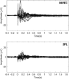
Figure 1
Figure 1. Grand average TMS-evoked potentials for both conditions (MPFC and SPL).
Zero corresponds to the TMS onset in each condition.
Figure 2
Figure 2. Grand average of the induced cortical alpha power after TMS on MPFC or SPL at t = 0.7 s with zero corresponding to the TMS onset.
The alpha power is represented as a percentage of ERS/ERD relative to the pre-TMS baseline (t = −0.4 s to −0.1 s). The star indicates the stimulated site.
Figure 3
Figure 3. Grand average occipital alpha power (αMPFC, αSPL) across time around the TMS pulse (occurring at t = 0).
The alpha power is represented as a percentage of ERS/ERD relative to the pre-TMS baseline (t = −0.4 s to −0.1 s). The mean basic alpha power during rest state period (αRest-state) for both conditions is represented as a dashed line. The annotated time periods mark:
  1. αSPL and αMPFC are not significantly different from αRest-state t = 0.28–0.45 s.

  2. αMPFC significantly higher than αSPL (p = 0.03) t = 0.78–1.44 s.

  3. αMPFC significantly higher than αRest-state (p = 0.0016) t = 0.48–2.07 s.

  4. αSPL significantly higher than αRest-state (p = 0.0207) t = 0.47–1.7 s.

Figure 4
Figure 4. For each stimulation condition, the targeted point is shown on MRI slices by the red dot.
An example of the induced electric field represented on a 3D reconstruction of the brain is also shown for each stimulated site, with the hand area in the primary motor cortex as a reference point. For left MPFC, we targeted the area BA10, i.e. anterior part of the superior frontal gyrus, very near the median line. The coil handle pointed backward with an angle of approximately 10° with respect to the midline. For left SPL, we targeted area BA7, i.e. part of the parietal cortex, located between the marginal branch of the cingulate sulcus and the parieto-occipital sulcus. The coil handle pointed towards the back of the head, forming a 45° angle with respect to the midline in the clockwise direction.

References

    1. Raichle M. E. et al.. A default mode of brain function. Proceedings of the National Academy of Sciences 98(2), 676–682 (2001). - PMC - PubMed
    1. Mantini D., Perrucci M. G., Del Gratta C., Romani G. L. & Corbetta M. Electrophysiological signatures of resting state networks in the human brain. Proceedings of the National Academy of Sciences 104(32), 13170–13175 (2007). - PMC - PubMed
    1. de Pasquale F. et al.. Temporal dynamics of spontaneous MEG activity in brain networks. Proc Natl Acad Sci USA 107(13), 6040–6045 (2010). - PMC - PubMed
    1. Chen A. C. et al.. Causal interactions between fronto-parietal central executive and default-mode networks in humans. Proc Natl Acad Sci USA 110(49), 19944–19949 (2013). - PMC - PubMed
    1. Bartolomeo P., Thiebaut de Schotten M. & Chica A. B. Brain networks of visuospatial attention and their disruption in visual neglect. Front Hum Neurosci 6, 110 (2012). - PMC - PubMed

Publication types

LinkOut - more resources