. 2016 Aug 3;36(31):8188-99.
doi: 10.1523/JNEUROSCI.3935-15.2016.

Feature-Selective Attentional Modulations in Human Frontoparietal Cortex

Affiliations

Feature-Selective Attentional Modulations in Human Frontoparietal Cortex

Edward F Ester et al. J Neurosci. .

Abstract

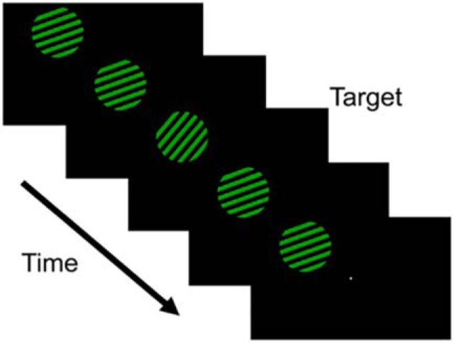
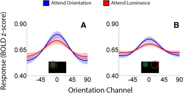
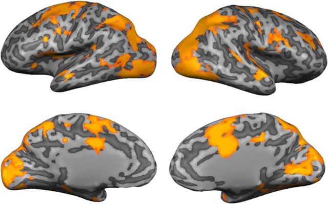
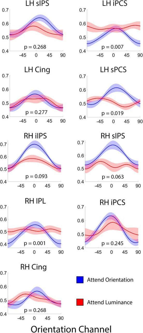
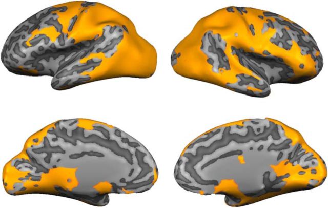
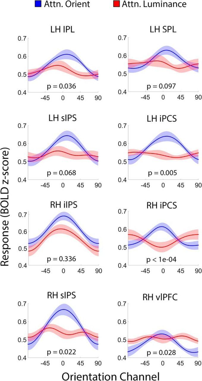
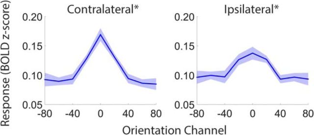
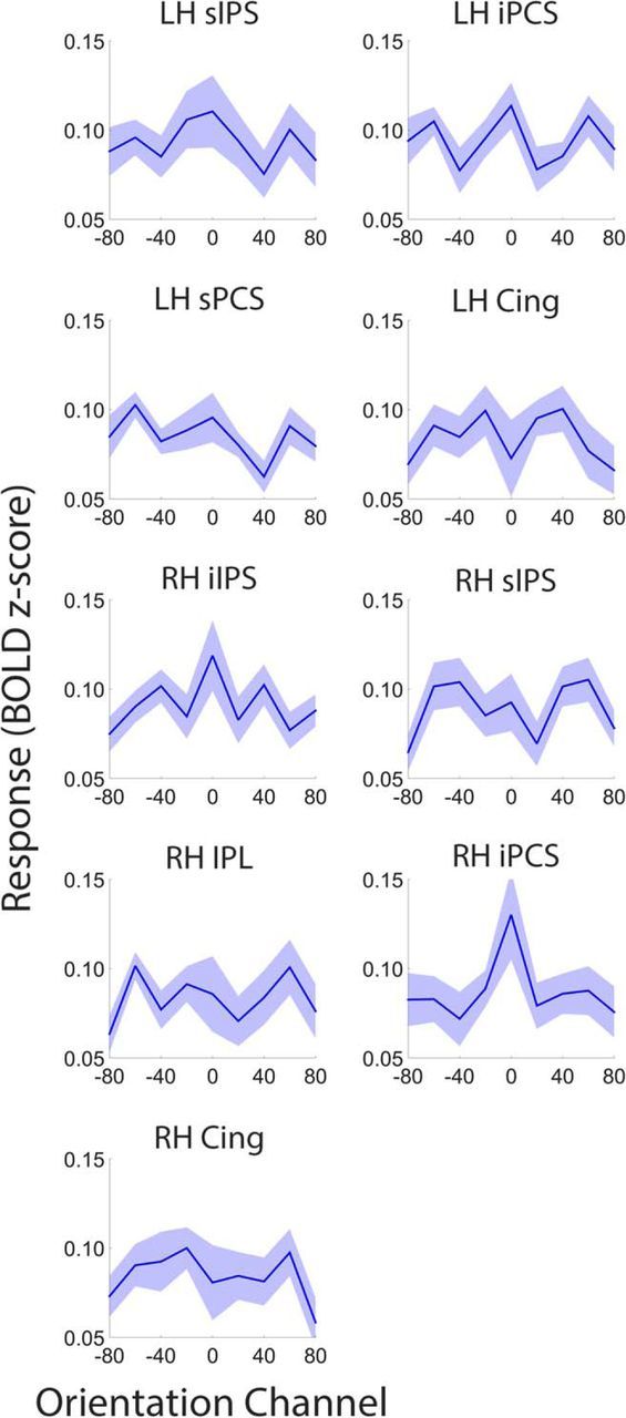
Control over visual selection has long been framed in terms of a dichotomy between "source" and "site," where top-down feedback signals originating in frontoparietal cortical areas modulate or bias sensory processing in posterior visual areas. This distinction is motivated in part by observations that frontoparietal cortical areas encode task-level variables (e.g., what stimulus is currently relevant or what motor outputs are appropriate), while posterior sensory areas encode continuous or analog feature representations. Here, we present evidence that challenges this distinction. We used fMRI, a roving searchlight analysis, and an inverted encoding model to examine representations of an elementary feature property (orientation) across the entire human cortical sheet while participants attended either the orientation or luminance of a peripheral grating. Orientation-selective representations were present in a multitude of visual, parietal, and prefrontal cortical areas, including portions of the medial occipital cortex, the lateral parietal cortex, and the superior precentral sulcus (thought to contain the human homolog of the macaque frontal eye fields). Additionally, representations in many-but not all-of these regions were stronger when participants were instructed to attend orientation relative to luminance. Collectively, these findings challenge models that posit a strict segregation between sources and sites of attentional control on the basis of representational properties by demonstrating that simple feature values are encoded by cortical regions throughout the visual processing hierarchy, and that representations in many of these areas are modulated by attention.

Significance statement: Influential models of visual attention posit a distinction between top-down control and bottom-up sensory processing networks. These models are motivated in part by demonstrations showing that frontoparietal cortical areas associated with top-down control represent abstract or categorical stimulus information, while visual areas encode parametric feature information. Here, we show that multivariate activity in human visual, parietal, and frontal cortical areas encode representations of a simple feature property (orientation). Moreover, representations in several (though not all) of these areas were modulated by feature-based attention in a similar fashion. These results provide an important challenge to models that posit dissociable top-down control and sensory processing networks on the basis of representational properties.

Keywords: frontoparietal cortex; functional neuroimaging; visual attention; visual cortex.


Figures

Figure 1.
Figure 1.
Stimulus displays. Participants viewed displays containing a single square-wave grating in the upper left or right visual field. On each trial, the grating was assigned one of nine orientations (0–160° in 20° increments) and one of two colors (yellow or green). In separate scans, participants were instructed to attend either the orientation or luminance of the grating. During attend-luminance scans, participants discriminated the direction (clockwise or anticlockwise) of brief and unpredictable perturbations in stimulus orientation. During attend-luminance scans, participants discriminated the direction (brighter or dimmer) of brief and unpredictable perturbations in stimulus luminance.
Figure 2.
Figure 2.
Representations of stimulus orientation in retinotopically organized visual cortex. Data have been averaged across visual areas V1, V2v, V3v, and hV4v and sorted by location relative to the stimulus on each trial (contralateral vs ipsilateral; A and B, respectively). Shaded areas are ±1 within-participant SEM.
Figure 3.
Figure 3.
Searchlight-defined ROIs encoding stimulus orientation. A leave-one-participant-out cross-validation scheme was used to generate an SPM of searchlight neighborhoods containing a robust representation of stimulus orientation for each participant (p < 0.01, FDR corrected for multiple comparisons; see Materials and Methods, Searchlight definition of ROIs representing stimulus orientation). Here, the SPMs for a representative participant (DM) have been projected onto a computationally inflated representation of his or her cortical sheet. For exposition, neighborhoods containing a robust representation of orientation have been assigned a value of 1 while neighborhoods that did not have been zeroed out. Across participants, robust representations of stimulus orientation were present in a broad network of visual, parietal, and frontal cortical areas (Table 1).
Figure 4.
Figure 4.
Reconstructed representations of orientation in frontoparietal cortex are modulated by task relevance. Each panel plots reconstructed representations of stimulus orientation measured during attend-orientation and attend-luminance scans in searchlight-defined ROIs that contained a robust representation of orientation (Table 1). The p value in each panel corresponds to the proportion of bootstrap permutations where amplitude estimates were reliably higher during attend-luminance relative to attend-orientation scans (FDR corrected for multiple comparisons); thus, a p value <0.05 indicates that amplitude estimates were reliably larger during attend-orientation scans relative to attend-luminance scans. Shaded regions are ±1 within-participant SEM. Cing, Cingulate gyrus; iIPS, inferior intraparietal sulcus; iPCS, inferior precentral sulcus; IPL, inferior parietal lobule; LH, left hemisphere; RH, right hemisphere; sIPS, superior intraparietal sulcus.
Figure 5.
Figure 5.
Searchlight-defined ROIs encoding task set. We combined a roving searchlight analysis with an SVM to identify cortical regions representing participants' task set (i.e., attend orientation vs attend luminance; p < 0.01, FDR corrected for multiple comparisons). Here, the resulting map has been projected onto a computationally inflated image of a representative participant's brain (DM). For exposition, searchlight neighborhoods containing a robust representation of orientation have been assigned a value of 1 while neighborhoods that did not have been zeroed out. From this map, we manually defined a set of 19 frontal, parietal, and inferior temporal ROIs that encoded task set (Table 2).
Figure 6.
Figure 6.
Attentional modulations in task-selective frontoparietal ROIs. Each panel plots reconstructed representations of stimulus orientation from searchlight-defined ROIs containing a robust representation of participants' task set (i.e., attend orientation or attend luminance; see Fig. 5 and Table 2). The p value in each panel corresponds to the proportion of bootstrap permutations where amplitude estimates were reliably higher during attend-luminance relative to attend-orientation scans (FDR corrected for multiple comparisons); thus, a p value <0.05 indicates that amplitude estimates were reliably larger during attend-orientation scans relative to attend-luminance scans. Shaded regions are ±1 within-participant SEM. iIPS, Inferior intraparietal sulcus; iPCS, inferior precentral sulcus; IPL, inferior parietal lobule; LH, left hemisphere; RH, right hemisphere; sIPS, superior intraparietal sulcus; SPL, superior parietal lobule; vlPFC, ventrolateral prefrontal cortex.
Figure 7.
Figure 7.
Continuous versus categorical representations in visual cortical ROIs. To examine whether the orientation-selective representations plotted in Figure 2 are continuous, we recomputed reconstructions of stimulus orientation from activation patterns measured in contralateral and ipsilateral visual areas during attend-orientation scans using a basis set of nonoverlapping delta functions. If the representation encoded by a given ROI is discrete or categorical, then the reconstructed representation computed using this approach should exhibit a sharp peak at the target orientation. We therefore computed the slope of the reconstructed representation in each ROI (see text for details). A p value < 0.05 indicates a positive slope and is consistent with a continuous rather than categorical or discrete representation. Shaded regions are ±1 within-participant SEM. *p < 0.05 and ∧p < 0.10, FDR corrected for multiple comparisons across ROIs.
Figure 8.
Figure 8.
Continuous versus categorical representations in searchlight amplitude ROIs. Compare with attend-orientation reconstructions in Figure 4. Conventions are in Figure 7. We were unable to reconstruct a representation of stimulus orientation in many ROIs. Shaded regions are ±1 within-participant SEM *p < 0.05 and ∧p < 0.10, FDR corrected for multiple comparisons across ROIs.
Figure 9.
Figure 9.
Continuous versus categorical representations in task-selective ROIs. Compare with attend-orientation reconstructions in Figure 6. Conventions are in Figures 7 and 8. Shaded regions are ±1 within-participant SEM. *p < 0.05 and ∧p < 0.10, FDR corrected for multiple comparisons across ROIs.

References

    1. Andersen SK, Hillyard SA, Müller MM. Attention facilitates multiple stimulus features in parallel in human visual cortex. Curr Biol. 2008;18:1006–1009. doi: 10.1016/j.cub.2008.06.030. - DOI - PubMed
    1. Baldauf D, Desimone R. Neural mechanisms of object-based attention. Science. 2014;344:424–427. doi: 10.1126/science.1247003. - DOI - PubMed
    1. Bichot NP, Heard MT, DeGennaro EM, Desimone R. A source for feature-based attention in the prefrontal cortex. Neuron. 2015;88:832–844. doi: 10.1016/j.neuron.2015.10.001. - DOI - PMC - PubMed
    1. Brainard D. The psychophysics toolbox. Spat Vis. 1997;10:433–436. doi: 10.1163/156856897x00357. - DOI - PubMed
    1. Bressler SL, Tang W, Sylvester CM, Shulman GL, Corbetta M. Top-down control of human visual cortex by frontal and parietal cortex in anticipatory visual spatial attention. J Neurosci. 2008;28:10056–10061. doi: 10.1523/JNEUROSCI.1776-08.2008. - DOI - PMC - PubMed

Publication types