. 2016 Aug 3;36(31):8228-37.
doi: 10.1523/JNEUROSCI.1244-16.2016.

Reciprocal Control of Drinking Behavior by Median Preoptic Neurons in Mice

Affiliations

Reciprocal Control of Drinking Behavior by Median Preoptic Neurons in Mice

Stephen B G Abbott et al. J Neurosci. .

Abstract

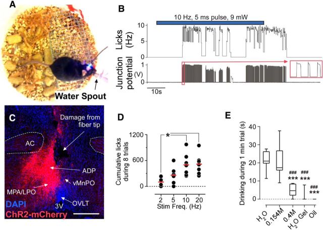
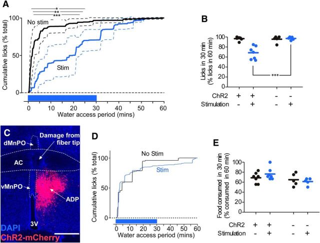
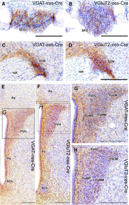
Stimulation of glutamatergic neurons in the subfornical organ drives drinking behavior, but the brain targets that mediate this response are not known. The densest target of subfornical axons is the anterior tip of the third ventricle, containing the median preoptic nucleus (MnPO) and organum vasculosum of the lamina terminalis (OVLT), a region that has also been implicated in fluid and electrolyte management. The neurochemical composition of this region is complex, containing both GABAergic and glutamatergic neurons, but the possible roles of these neurons in drinking responses have not been addressed. In mice, we show that optogenetic stimulation of glutamatergic neurons in MnPO/OVLT drives voracious water consumption, and that optogenetic stimulation of GABAergic neurons in the same region selectively reduces water consumption. Both populations of neurons have extensive projections to overlapping regions of the thalamus, hypothalamus, and hindbrain that are much more extensive than those from the subfornical organ, suggesting that the MnPO/OVLT serves as a key link in regulating drinking responses.

Significance statement: Neurons in the median preoptic nucleus (MnPO) and organum vasculosum of the lamina terminalis (OVLT) are known to regulate fluid/electrolyte homeostasis, but few studies have examined this issue with an appreciation for the neurochemical heterogeneity of these nuclei. Using Cre-Lox genetic targeting of Channelrhodospin-2 in transgenic mice, we demonstrate that glutamate and GABA neurons in the MnPO/OVLT reciprocally regulate water consumption. Stimulating glutamatergic MnPO/OVLT neurons induced water consumption, whereas stimulating GABAergic MnPO neurons caused a sustained and specific reduction in water consumption in dehydrated mice, the latter highlighting a heretofore unappreciated role of GABAergic MnPO neurons in thirst regulation. These observations represent an important advance in our understanding of the neural circuits involved in the regulation of fluid/electrolyte homeostasis.

Keywords: VGAT; VGLUT2; dehydration; lamina terminalis; optogenetics; thirst.


Figures

Figure 1.
Figure 1.
Distribution of VGluT2 and VGAT neurons in the POA. A, B, Mid-sagittal section showing the dorsal MnPO (dMnPO) and ventral MnPO (vMnPO), and OVLT in Slc17a6-cre;Rpl10-GFP (i.e., VGluT2-ires-Cre-GFP; A) and Slc32a1-cre;Rpl10-GFP (i.e., VGAT-ires-Cre-GFP; B) mice. C, D, Anterior POA coronal section at bregma + 0.30 mm showing that the density of glutamatergic neurons is higher in the MnPO than surrounding areas, whereas the reverse is true for GABAergic neurons. E, F, Sagittal schematic for injections of AAV-FLEX-ChR2-mCherry showing the angle of approach (E) and the tract left by optical fiber for stimulation of the MnPO/OVLT (F). G, H, Examples of injection sites that hit the MnPO, visualized by the expression of mCherry (native fluorescence). Note that, due to the low density of glutamatergic neurons outside the MnPO, the injection site in a VGluT2-ires-CreChR2+ animal is largely confined to the MnPO, but in a VGAT-ires-CreChR2+ animal, numerous cells outside the MnPO express ChR2-mCherry. Scale bars: A, B, 200 μm; C, D, 250 μm; F, 1.5 mm; G, H, 250 μm. ac, Anterior commissure; cc, corpus callosum; f, fornix; HDB, nucleus of the horizontal limb of the diagonal band; ox, optic chiasm; Pe, periventricular preoptic nucleus; 3 V, third ventricle.
Figure 2.
Figure 2.
Fiber optic placement and location of neurons expressing ChR2-mcherry in experimental cases. A–D, Optical fiber tip placement, and distribution of ChR2-mcherry neurons in VGluT2-ires-CreChR2 (N = 7; A), VGluT2-ires-CreChR2+ (N = 8; B), VGAT-ires-CreChR2 (N = 5; C), and VGAT-ires-CreChR2+ (N = 7; D) mice. Bregma level is identified on the far right.
Figure 3.
Figure 3.
Stimulating excitatory neurons in the MnPO/OVLT cause water consumption. A, Experimental setup for testing optogenetic driving of drinking behavior. B, Raw data from lickometer showing onset and pattern of drinking during 1 min stimulation (10 Hz, 5 ms pulse, 11 mW). C, An injection site in mouse id224 (VGluT2-ires-CreChR2+), in which the injection was lateral to the MnPO and the animal did not consume water during stimulation. Bregma level, +0.42 mm. Scale bars, 250 μm. MPA/LPO, Medial/lateral preoptic area. D, Total number of spout licks during a series of eight 1 min trials (i.e., 1 min on/1 min off). *p < 0.05. E, Solution preference assay (N = 7; ***p < 0.001 vs H2O, ###p < 0.001 vs saline). Box-and-whisker plot reflects range, 25% and 75% range, and median.
Figure 4.
Figure 4.
Stimulating inhibitory neurons in the MnPO/OVLT reduces water consumption, but not food consumption. A, Normalized cumulative spout licks (mean ± SD, N = 7) during 60 min of water access in water-deprived VGAT-ires-creChR2+ mice. Stimulation (1 s on/2 s off, 20 Hz, 5 ms, 13 mW; in blue) was applied for the first 30 min of the water access period. *p < 0.05, **p < 0.01, ***p < 0.001 by post hoc t test with Bonferroni's correction. B, Normalized spout licks by water-deprived mice during the first 30 min of the 60 min access period. (***p < 0.001 by post hoc t test with Bonferroni's correction). C, Injection site in a VGAT-ires-CreChR2+ mouse in which the injection was lateral to the MnPO, and in which the animal had no response to optogenetic stimulation during water repletion after dehydration. Bregma level, −0.12 mm. Scale bars, 250 μm. D, Time course of cumulative water spout licks during a 1 h access period following overnight water deprivation for the mouse shown in C. E, Food consumed by food-deprived mice during the first 30 min of food access as a percentage of the total amount of food consumed in 60 min.
Figure 5.
Figure 5.
Projections of VGluT2 and VGAT neurons. A–J, Glutamatergic and GABAergic MnPO/OVLT neurons innervate the paraventricular thalamic nucleus (A–D); the lateral habenula (C, D); the tuberal hypothalamus (E, F); the periaqueductal gray matter, raphe nuclei, and pedunculopontine tegmental region (G, H); laterodorsal tegmental nucleus, locus ceruleus, and Barrington's nucleus (I, J); and the medullary raphe (K, L). Injection sites for each respective case are shown in Figure 1, G and H. Scale bar, 200 μm. AM, Anterodorsal thalamic nucleus; Arc, arcuate nucleus; Bar, Barrington's nucleus; dPAG, dorsal periaqueductal gray; DMH, dorsomedial hypothalamus; DR, dorsal raphe; DTg, dorsal tegmentum; f, fornix; me5, mesenphalic nucleus 5; MHB, medial habenula; mlf, medial longitudinal fasciculus; MnR, median raphe; PPT, pedunculopontine tegmental nucleus; LC, locus coeruleus; LDT, lateral dorsal tegmentum; LHA, lateral hypothalamic area; LHB, lateral habenula; Pe, periventricular area; PV, paraventricular nucleus of the thalamus; py, pyramidal tract; Re, reuniens thalamic nucleus; RMg, raphe magnus; RPa, raphe pallidus; vlPAG, ventrolateral periaqueductal gray; VMH, ventromedial hypothalamus; xscp, decussation of the superior cerebellar peduncle; ZI, zona incerta.
Figure 6.
Figure 6.
Projections to MnPO/OVLT VGluT2 and VGAT neurons in brain regions involved in neuroendocrine response to dehydration. A–H, Glutamatergic and GABAergic MnPO/OVLT neurons innervate the subfornical organ (A, B) supraoptic nucleus (C, D), and paraventricular nucleus of the hypothalamus (E–H). Injection sites for each respective case are shown in Figure 1, G and H. Scale bar, 200 μm. opt, Optic tract; PaDC, paraventricular hypothalamic nucleus (dorsal cap); PaLM, paraventricular hypothalamic nucleus (lateral magnocellular); PaMM, paraventricular hypothalamic nucleus (medial magnocellular); PaMP, paraventricular hypothalamic nucleus (medial parvicellular); Pe, periventricular hypothalamic nucleus; Re, reuniens thalamic nucleus; SCh, suprachiasmatic nucleus.

Similar articles

Cited by

References

    1. Bourque CW. Central mechanisms of osmosensation and systemic osmoregulation. Nat Rev Neurosci. 2008;9:519–531. doi: 10.1038/nrn2400. - DOI - PubMed
    1. Buggy J, Johnson AK. Anteroventral third ventricle periventricular ablation: temporary adipsia and persisting thirst deficits. Neurosci Lett. 1977;5:177–182. doi: 10.1016/0304-3940(77)90043-X. - DOI - PubMed
    1. Cunningham JT, Sullivan MJ, Edwards GL, Farinpour R, Beltz TG, Johnson AK. Dissociation of experimentally induced drinking behavior by ibotenate injection into the median preoptic nucleus. Brain Res. 1991;554:153–158. doi: 10.1016/0006-8993(91)90183-V. - DOI - PubMed
    1. Cunningham JT, Beltz T, Johnson RF, Johnson AK. The effects of ibotenate lesions of the median preoptic nucleus on experimentally-induced and circadian drinking behavior in rats. Brain Res. 1992;580:325–330. doi: 10.1016/0006-8993(92)90961-8. - DOI - PubMed
    1. De Luca LA, Jr, Xu Z, Schoorlemmer GH, Thunhorst RL, Beltz TG, Menani JV, Johnson AK. Water deprivation-induced sodium appetite: humoral and cardiovascular mediators and immediate early genes. Am J Physiol Regul Integr Comp Physiol. 2002;282:R552–R559. doi: 10.1152/ajpregu.00295.2000. - DOI - PubMed

Publication types