

Pigmented anatomy in Carboniferous cyclostomes and the evolution of the vertebrate eye
- PMID: 27488650
- PMCID: PMC5013770
- DOI: 10.1098/rspb.2016.1151
Pigmented anatomy in Carboniferous cyclostomes and the evolution of the vertebrate eye
Abstract
The success of vertebrates is linked to the evolution of a camera-style eye and sophisticated visual system. In the absence of useful data from fossils, scenarios for evolutionary assembly of the vertebrate eye have been based necessarily on evidence from development, molecular genetics and comparative anatomy in living vertebrates. Unfortunately, steps in the transition from a light-sensitive 'eye spot' in invertebrate chordates to an image-forming camera-style eye in jawed vertebrates are constrained only by hagfish and lampreys (cyclostomes), which are interpreted to reflect either an intermediate or degenerate condition. Here, we report-based on evidence of size, shape, preservation mode and localized occurrence-the presence of melanosomes (pigment-bearing organelles) in fossil cyclostome eyes. Time of flight secondary ion mass spectrometry analyses reveal secondary ions with a relative intensity characteristic of melanin as revealed through principal components analyses. Our data support the hypotheses that extant hagfish eyes are degenerate, not rudimentary, that cyclostomes are monophyletic, and that the ancestral vertebrate had a functional visual system. We also demonstrate integument pigmentation in fossil lampreys, opening up the exciting possibility of investigating colour patterning in Palaeozoic vertebrates. The examples we report add to the record of melanosome preservation in Carboniferous fossils and attest to surprising durability of melanosomes and biomolecular melanin.
Keywords: Mazon Creek; cyclostomes; melanosomes; retinal pigment epithelium.
© 2016 The Authors.
Figures


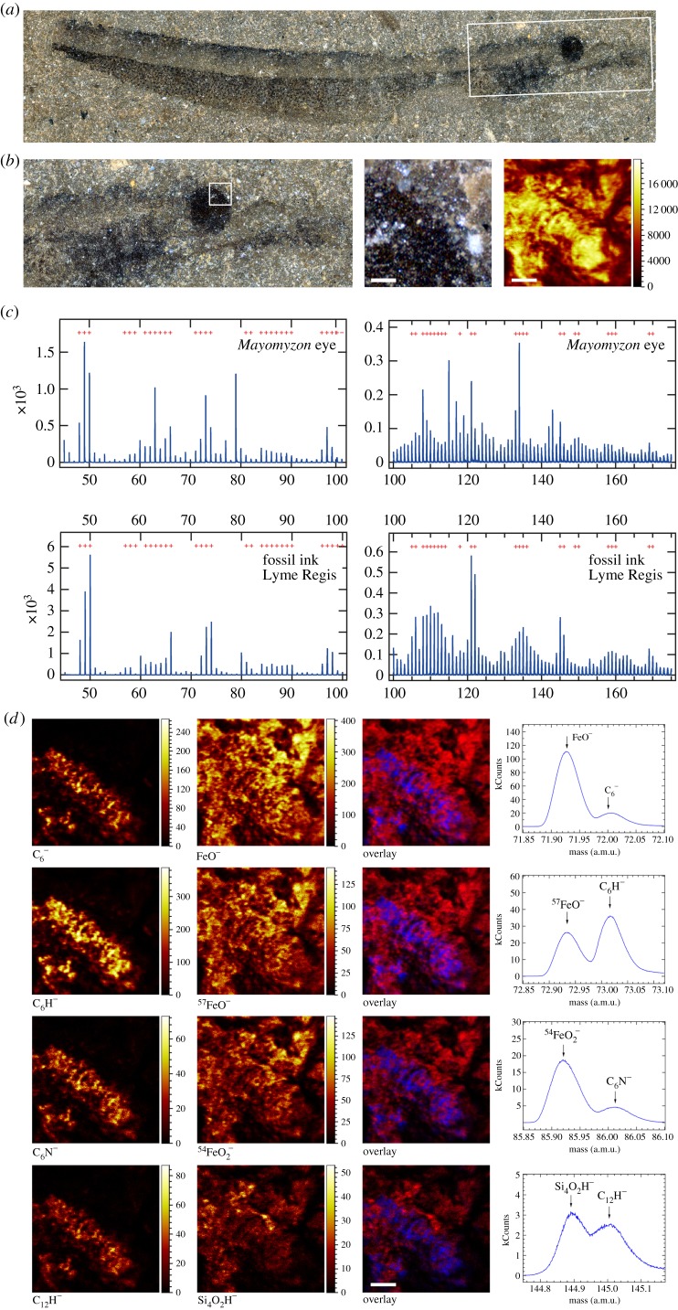
 , C6H−, C6N− and C12H−, together with matrix-related fragments: FeO−, 57FeO−,
  and Si4O2H−. Scale bar, 100 µm. See also the electronic supplementary material, figures S4 and S6 for additional maps and spectra. (Online version in colour.)
References
-
- Locket NA, Jørgensen JM. 1998. The eyes of hagfishes. In The biology of hagfishes (eds JM Jørgensen, JP Lomholt, RE Weber, H Malte), pp. 541–556. Berlin, Germany: Springer.
MeSH terms
Associated data
LinkOut - more resources
Full Text Sources
Other Literature Sources