. 2016 Aug 4:7:12259.
doi: 10.1038/ncomms12259.

Load-induced enhancement of Dynein force production by LIS1-NudE in vivo and in vitro

Affiliations

Load-induced enhancement of Dynein force production by LIS1-NudE in vivo and in vitro

Babu J N Reddy et al. Nat Commun. .

Abstract

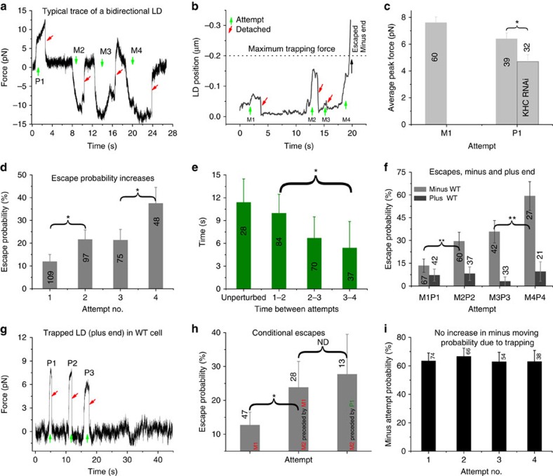
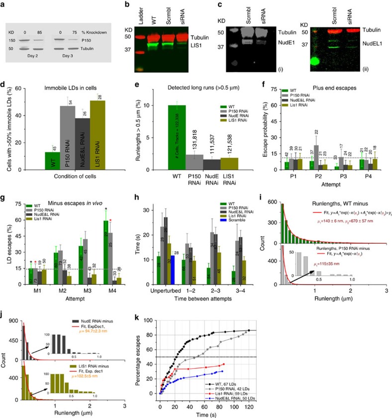
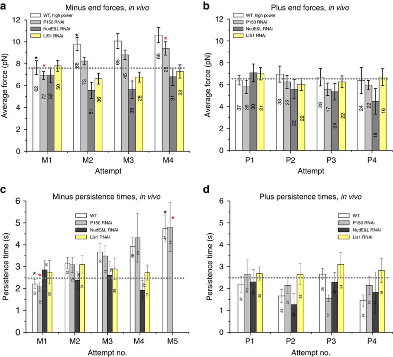
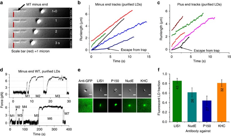
Most sub-cellular cargos are transported along microtubules by kinesin and dynein molecular motors, but how transport is regulated is not well understood. It is unknown whether local control is possible, for example, by changes in specific cargo-associated motor behaviour to react to impediments. Here we discover that microtubule-associated lipid droplets (LDs) in COS1 cells respond to an optical trap with a remarkable enhancement in sustained force production. This effect is observed only for microtubule minus-end-moving LDs. It is specifically blocked by RNAi for the cytoplasmic dynein regulators LIS1 and NudE/L (Nde1/Ndel1), but not for the dynactin p150(Glued) subunit. It can be completely replicated using cell-free preparations of purified LDs, where duration of LD force production is more than doubled. These results identify a novel, intrinsic, cargo-associated mechanism for dynein-mediated force adaptation, which should markedly improve the ability of motor-driven cargoes to overcome subcellular obstacles.


Figures

Figure 1
Figure 1. Force adaptation occurs for lipid droplets (LDs) in vivo in the minus end direction.
(a) Typical trace of LD escaping the optical trap. In the figures, ‘M#' and ‘P#' denote direction and escape attempt # of the LD in laser trap, that is, the first escape attempts towards the minus and plus end of the microtubule are denoted by ‘M1' and ‘P1', respectively. Unless mentioned otherwise, the numbers in/above the bars in the figures hereafter are the N values for the measurements. (b) High resolution bi-directional force trace of a LD showing increased minus-end force persistence and higher force with time. (c) Average peak plus end forces decrease due to KHC siRNA treatment (data shown is from 2 different sets of cultures from ∼25 cells and reduced forces were observed in four trials). (d) Probability of LDs escaping from the laser trap increases with attempt number. (e) Average time between periods of linear motion (Unperturbed) or attempts (when trapped) of LDs in WT cells. (f) Percentage of LDs escaping from the trap in the minus- and plus-end directions as a function of attempt number. (g) Trace showing lack of adaptation in the plus end moving LD in the trap. (h) Minus-end adaptation occurs even when a previous (failed) attempt was in the plus-end direction. (i) No change in the probability of a given attempt occurring in the minus end direction as a function of attempt #. (In dh, trajectories of 109 LDs were analysed from cells cultured on four different days. At least five cells were analysed in each dish lasting for about an hour. See methods. Overall, force persistence adaptation of LDs was observed in more than 20 different sets of WT cultures.) *P<0.05, **P<0.01, t-test Error bars=s.e.m.
Figure 2
Figure 2. NudE and L/LIS1 contribute to force escape adaptation and dynactin to on-rate adaption.
(ac) Western blots quantifying the levels of P150, LIS1 and NudE(i)& NudEL(ii) in control and siRNA treated cells. (d) Inside COS1 cells, when LIS1, NudE and L and P150 levels are reduced, there is an increase in immobile LDs. N values indicate no. of cells (e) In siRNA treated cells, there are fewer long runs (>0.5 microns) compared to the WT. (f) Escape percentages in the plus end direction in LIS1, NudE and L and P150 SiRNA treated cells are similar to WT cells. The apparent larger escape probability for P2/P150 is statistically significant but unlikely to be real (Supplementary Note 4) (g) Minus end force adaptation is absent in the cells with low levels of LIS1 and NudE and L, but still occurs in the reduced P150 background (h) In the decreased P150 background, the time between attempts is longer and does not adapt as it does in the WT. Lack of attempt-frequency adaptation also occurs in the NudE and LIS1 knockdowns. WT data in 2h reflects additional control measurements made simultaneously with the RNAi knockdown measurements, and is thus independent of data presented in Fig 1e. (i) Run lengths are decreased in P150 siRNA cells (bottom) relative to the wild type (top). Similarly in NudE/L and LIS1 siRNA cells, LDs have shorter runs(j). (k)Cumulative percentage of LD population that escaped from the optical trap in vivo, as a function of time in WT, P150, LIS1, and NudE and L SiRNA cells. Error bars=s.e.m. In f and g they are proportional errors. *P<0.05 t-test. Reported siRNA phenotypes were observed in at least 5 different days of cultures and at least 20 cells were analysed per set.
Figure 3
Figure 3. High power measurements in WT cells show LD adaptation reflects increased force persistence
(a) The average peak forces of all LDs in the minus end direction shows a slight increase for WT and P150siRNA cells but not for LIS1 and NudE&L siRNA treated cells. (b) The average peak forces of all LDs in the plus end direction does not change. (c,d) In WT and P150 siRNA cells, the average persistence time of all attempts increases significantly in the minus end, but not in the plus end, direction. (99 LDs, excluding measurements in Fig. 1, were analysed for WT using high power trap to minimize the escapes). The average persistence time of minus-end LD attempts does not increase in LIS1 or NudE and L siRNA treated cells in any direction. Error bars=s.e.m. *P<0.05, t-test.
Figure 4
Figure 4. Reconstitution of LD motion and adaptation in vitro.
(a) Purified LD moving towards minus end of polarity marked microtubule (Minus ends are small biotinylated MTs attached with beads prepared as mentioned in Soppina et al., (supplement) excepting that beads used are streptavidin coated,150 nm MagCellect from R&D Systems, USA). (b,c) Typical tracks of purified LDs in the minus and plus end direction respectively. (d) Typical minus end trace of purified LD. Note that the lower trace in d has a low (atypical) on-rate, but was chosen because it includes a few small attempts (M1 and M3) as well as an extremely long duration event (M6). (e) DIC and TIRF images of LDs immuno-stained for LIS1, NudE, P150 and KHC proteins (also see Supplementary Fig. 5). The first panel is the GFP control showing no signal. Control GFP reflects use of a primary anti-GFP antibody (no GFP-labelled proteins present) made in rabbit (same host as the LIS1ab), to confirm that the LIS1 signal was not due to nonspecific binding of anti-LIS1ab. Scale bar=1 μm. (f) Fraction of LDs showing the presence of LIS1, NudE, P150 and KHC in the in vitro immuno-staining experiments. Error bars=Binomial standard error of proportion.
Figure 5
Figure 5. LD motion and adaptation in vitro.
(a,b) On-rates and persistence times of motors on purified LDs moving in the minus and plus ends are much higher (top traces) compared to the same in cells (bottom traces). (c) Motile fraction of purified LDs moving along polarity-marked taxol-stabilized microtubules (∼22%) is slightly larger than that of LDs moving in vivo (∼5–10%) at any given instant. (d,e ) Average forces of WT purified LDs increase slightly in both the directions. Both LIS1 and NudE function blocking antibodies and the NudE fragment reduced the forces. (f,g) Force persistence times for WT LDs are much higher than in vivo in both directions. As in vivo, persistence durations adapt and increase in the minus-end direction. Function blocking antibodies to LIS1 and NudE eliminated the adaptation, as did NudE fragment (10-191 aa). (h,i). Typical traces of Minus end moving LDs illustrating lower forces in the presence of function blocking abs to LIS1 and NudE while the plus end motion (j) is unaffected.(k)Typical traces of NudE191 treated LDs show relatively short persistence times(compare them to top panels in a,b). For de, labels in M1 and P1 indicate number of LDs tested. LD motion was observed in more than 10 different purifications. *P<0.05 t-test. #Not significantly different. Error bars are s.e.m. for dg and proportional errors for c.
Figure 6
Figure 6. Model.
(a) Force persistence model to explain the increase in LD escape probability (1) Dynein-NudE-LIS1(DNL) in the unadapted state (2) DNL under high load, after a conformational change in NudE positions LIS1 to interact with the dynein heads allowing it to change their MT detachment dynamics (b) Comparison of experiment and theoretical simulations assuming the presence of 13 dynein motors. (c,d) Average peak force and persistence times from persistence model before and after adaptation agree well with experiment. (e) Escape probabilities of trapped LDs from in vivo agree well with simulated escapes, using switching/persistence model; number of active DNL complexes assumed as indicated. *P<0.05, t-test. Error bars are s.e.m. in c,d and proportional errors in e.

Comment in

  • Dynein: Let's not get stuck!
    Reddy BJ, Gross S. Reddy BJ, et al. Cell Cycle. 2017 Jan 2;16(1):7-8. doi: 10.1080/15384101.2016.1232085. Epub 2016 Sep 22. Cell Cycle. 2017. PMID: 27657557 Free PMC article. No abstract available.

References

    1. Shubeita G. T. et al.. Consequences of motor copy number on the intracellular transport of kinesin-1-driven lipid droplets. Cell 135, 1098–1107 (2008). - PMC - PubMed
    1. Blehm B. H., Schroer T. A., Trybus K. M., Chemla Y. R. & Selvin P. R. In vivo optical trapping indicates kinesin's stall force is reduced by dynein during intracellular transport (vol 110, pg 3381, 2013). Proc. Natl Acad. Sci. USA 110, 9613–9613 (2013). - PMC - PubMed
    1. Fu M. M. & Holzbaur E. L. JIP1 regulates the directionality of APP axonal transport by coordinating kinesin and dynein motors. J. Cell Biol. 202, 495–508 (2013). - PMC - PubMed
    1. Yi J. Y. et al.. High-resolution imaging reveals indirect coordination of opposite motors and a role for LIS1 in high-load axonal transport. J. Cell Biol. 195, 193–201 (2011). - PMC - PubMed
    1. Coleman M. Axon degeneration mechanisms: commonality amid diversity. Nat. Rev. Neurosci. 6, 889–898 (2005). - PubMed

Publication types