. 2016 Sep 29;90(20):9182-93.
doi: 10.1128/JVI.01399-16. Print 2016 Oct 15.

Neuroinvasion of α-Synuclein Prionoids after Intraperitoneal and Intraglossal Inoculation

Affiliations

Neuroinvasion of α-Synuclein Prionoids after Intraperitoneal and Intraglossal Inoculation

Sara Breid et al. J Virol. .

Abstract

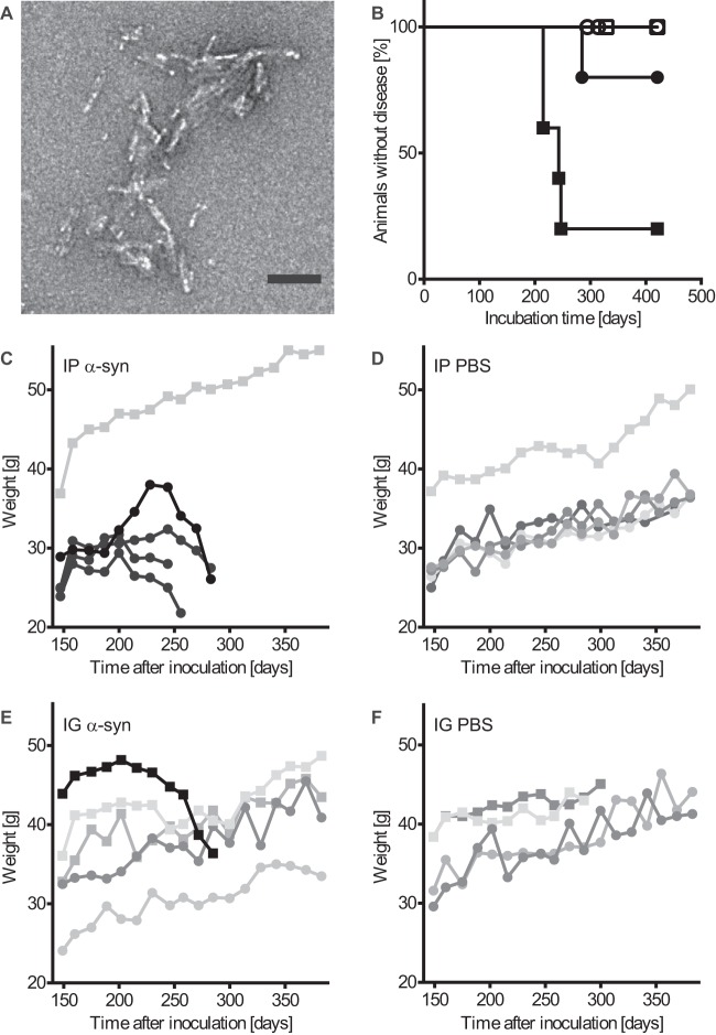
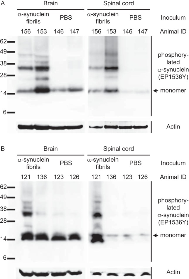
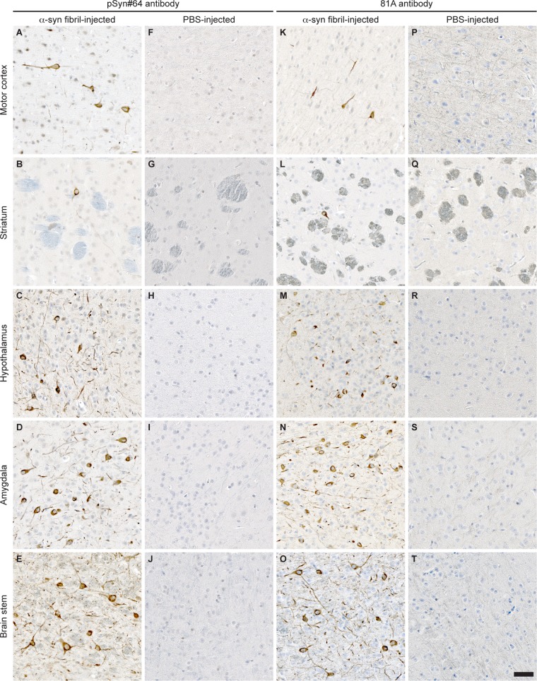
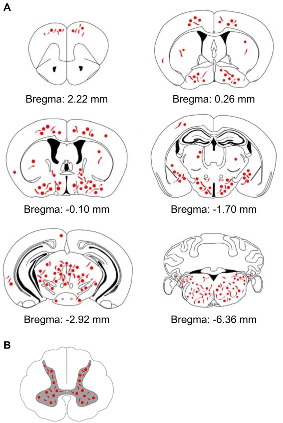
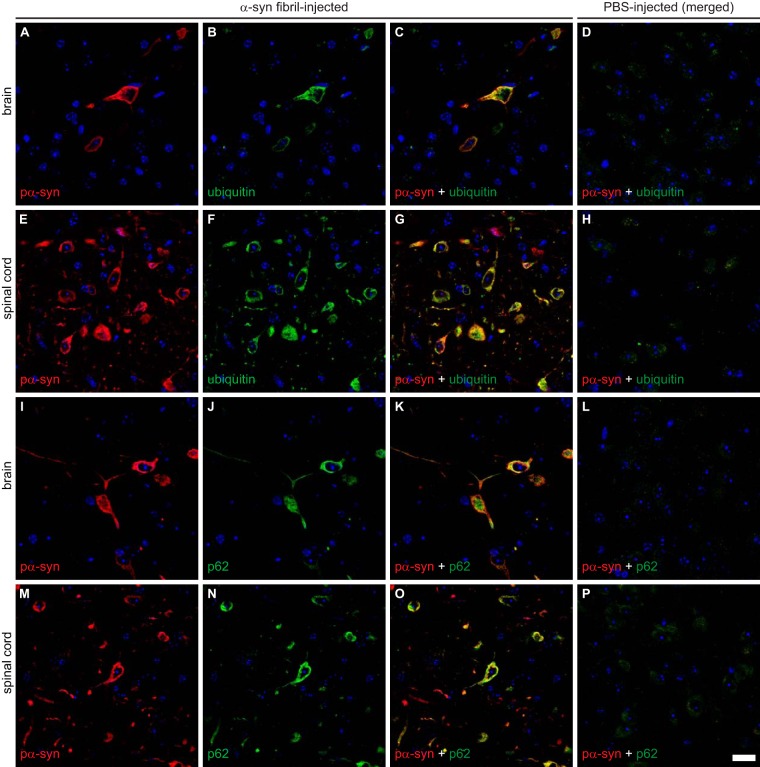
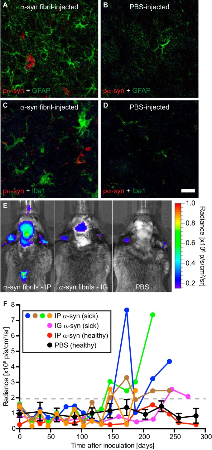
α-Synuclein is a soluble, cellular protein that in a number of neurodegenerative diseases, including Parkinson's disease and multiple system atrophy, forms pathological deposits of protein aggregates. Because misfolded α-synuclein has some characteristics that resemble those of prions, we investigated its potential to induce disease after intraperitoneal or intraglossal challenge injection into bigenic Tg(M83(+/-):Gfap-luc(+/-)) mice, which express the A53T mutant of human α-synuclein and firefly luciferase. After a single intraperitoneal injection with α-synuclein fibrils, four of five mice developed paralysis and α-synuclein pathology in the central nervous system, with a median incubation time of 229 ± 17 days. Diseased mice accumulated aggregates of Sarkosyl-insoluble and phosphorylated α-synuclein in the brain and spinal cord, which colocalized with ubiquitin and p62 and were accompanied by gliosis. In contrast, only one of five mice developed α-synuclein pathology in the central nervous system after intraglossal injection with α-synuclein fibrils, after 285 days. These findings are novel and important because they show that, similar to prions, α-synuclein prionoids can neuroinvade the central nervous system after intraperitoneal or intraglossal injection and can cause neuropathology and disease.

Importance: Synucleinopathies are neurodegenerative diseases that are characterized by the pathological presence of aggregated α-synuclein in cells of the nervous system. Previous studies have shown that α-synuclein aggregates made of recombinant protein or derived from brains of patients can spread in the central nervous system in a spatiotemporal manner when inoculated into the brains of animals and can induce pathology and neurologic disease, suggesting that misfolded α-synuclein can behave similarly to prions. Here we show that α-synuclein inoculation into the peritoneal cavity or the tongue in mice overexpressing α-synuclein causes neurodegeneration after neuroinvasion from the periphery, which further corroborates the prionoid character of misfolded α-synuclein.


Figures

FIG 1
FIG 1
Challenge with α-synuclein fibrils causes disease in bigenic Tg(M83+/−:Gfap-luc+/−) mice. (A) Negatively stained electron micrograph of sonicated recombinant human α-synuclein fibrils that were injected into mice. Aggregated samples were stained with uranyl acetate. Multiple clusters of short, rod-shaped aggregates are visible. Bar = 100 nm. (B) Kaplan-Meier survival curves show that after intraperitoneal injection with α-synuclein fibrils, four of five Tg(M83+/−:Gfap-luc+/−) mice developed signs of neurologic dysfunction in 229 ± 17 days (black squares), whereas none of the PBS-injected control mice developed neurologic disease within 420 days (white squares). One mouse of five died 285 days after intraglossal inoculation with α-synuclein fibrils (black circles). In contrast, none of the PBS-injected mice developed neurologic disease or spontaneously died (white circles). (C) Four (black symbols) of five mice that had been inoculated intraperitoneally (IP) with α-synuclein (α-syn) fibrils continuously lost weight for 6 to 8 weeks before they developed signs of neurologic disease. The one surviving animal continuously gained weight and did not develop disease or pathology for up to 420 days after inoculation (gray symbols). (E) One (black symbols) of five mice inoculated intraglossally (IG) with α-synuclein fibrils continuously lost weight for at least 8 weeks before it died. The four mice that did not succumb to disease or die continued to gain weight (gray symbols). Mice injected intraperitoneally (D) or intraglossally (F) with PBS gained weight throughout the course of the experiment. In panels C to F, male mice are represented by squares and female mice by circles.
FIG 2
FIG 2
Biochemical analysis of phosphorylated α-synuclein in the CNS of peripherally injected Tg(M83+/−:Gfap-luc+/−) mice. (A) Detection with the EP1536Y antibody, which recognizes phosphorylation at Ser129 of α-synuclein, showed that diseased mice accumulated high-molecular-weight species of Sarkosyl-insoluble aggregates of phosphorylated α-synuclein in their brains and spinal cords, whereas control mice challenged with PBS did not and showed bands only for the monomeric form of phosphorylated α-synuclein. (B) After intraglossal challenge with α-synuclein fibrils, only one mouse, animal 121, accumulated Sarkosyl-insoluble aggregates of phosphorylated α-synuclein in the brain and spinal cord, whereas all other intraglossally inoculated animals remained healthy. Molecular masses are shown in kilodaltons. Sample loading in each lane is shown by detection of actin.
FIG 3
FIG 3
Immunohistochemical analysis shows neuropathology in brains of Tg(M83+/−:Gfap-luc+/−) mice after intraperitoneal injection with α-synuclein fibrils. (A to E and K to O) Brain sections from animals injected with α-synuclein fibrils accumulated deposits of phosphorylated α-synuclein in multiple brain regions, as detected with the pSyn#64 antibody (A to E) and the 81A antibody (K to O), which recognize phosphorylation at Ser129 of α-synuclein. (F to J and P to T) In contrast, none of the PBS-injected mice showed any detectable aggregates of phosphorylated α-synuclein in the brain. Bar = 50 μm.
FIG 4
FIG 4
Immunohistochemistry shows abnormal, phosphorylated α-synuclein deposits in spinal cords of Tg(M83+/−:Gfap-luc+/−) mice after intraperitoneal injection with α-synuclein fibrils. (A) Deposits of phosphorylated α-synuclein were present in spinal cord sections from diseased animals, as illustrated by staining with the pSyn#64 antibody. (C and E) These deposits were composed of transgenically expressed human A53T α-synuclein, as shown with the Syn211 antibody (C), and endogenously expressed mouse α-synuclein, as visualized with the D37A6 antibody (E). (B, D, and F) None of the PBS-injected animals accumulated deposits of aggregated α-synuclein in the spinal cord. Bar = 50 μm.
FIG 5
FIG 5
Schematic overview showing the distribution of deposits of phosphorylated α-synuclein in the CNS of diseased Tg(M83+/−:Gfap-luc+/−) mice after intraperitoneal challenge with α-synuclein fibrils. (A) The distributions of abnormal deposits detected by use of two different antibodies, pSyn#64 and 81A, were comparable, and deposits could be found almost throughout the brain. However, the cerebellum was devoid of abnormal deposits of phosphorylated α-synuclein. (B) Abnormal aggregates of phosphorylated α-synuclein were also present in neurons within the gray matter of the spinal cords of diseased animals.
FIG 6
FIG 6
Phosphorylated α-synuclein can be detected in motor neurons of diseased Tg(M83+/−:Gfap-luc+/−) mice. (A to C) Immunofluorescence analysis showed that deposits of phosphorylated α-synuclein, as stained with the pSyn#64 antibody, were widespread in the gray matter and could also be detected in motor neurons within the ventral horn of the spinal cord in diseased Tg(M83+/−:Gfap-luc+/−) mice. (D to F) PBS-injected healthy control animals did not show aggregates of phosphorylated α-synuclein in their motor neurons. Motor neurons were detected with an antibody to choline O-acetyltransferase (ChAT). Nuclear staining with DAPI is shown in blue. Bar = 20 μm.
FIG 7
FIG 7
Immunofluorescence analysis shows that deposits of phosphorylated α-synuclein colocalize with ubiquitin and p62 in the brains and spinal cords of diseased Tg(M83+/−:Gfap-luc+/−) mice. Phosphorylated α-synuclein, detected with the EP1536Y antibody, and ubiquitin colocalized in the brains (A to C) and spinal cords (E to G) of fibril-injected diseased mice. Similarly, phosphorylated α-synuclein, detected here with the pSyn#64 antibody, also colocalized with p62 in brains (I to K) and spinal cords (M to O) of diseased mice. (D, H, L, and P) PBS-injected healthy control animals did not show aggregation or colocalization for any of these proteins. Nuclear staining with DAPI is shown in blue. Bar = 20 μm.
FIG 8
FIG 8
Gliosis in the brains of diseased Tg(M83+/−:Gfap-luc+/−) mice. (A) Immunofluorescence analysis of brain sections with an antibody against GFAP, a marker of astrocytes, showed that diseased animals had reactive astrogliosis in areas with deposits of phosphorylated α-synuclein, which were detected in the brain stem by use of the pSyn#64 antibody. (B) In contrast, reactive astrogliosis was not observed in brains of healthy, PBS-injected control mice. (C and D) Similarly, staining with an antibody to IBA-1, a marker of microglia, showed that diseased animals had microgliosis in areas with deposits of phosphorylated α-synuclein (C), which was not observed in brains of healthy, PBS-injected control mice (D). (E) Bioluminescence imaging revealed that Tg(M83+/−:Gfap-luc+/−) mice that had been challenged intraperitoneally with α-synuclein fibrils showed elevated radiance from their brains and spinal cords (left panel), caused by the increased activation of astrocytes, shortly before they developed neurologic symptoms, but this was not observed in PBS-injected control mice (right panel). After intraglossal inoculation with α-synuclein fibrils, one Tg(M83+/−:Gfap-luc+/−) mouse, animal 121, showed signs of reactive astrogliosis shortly before it died, at 285 days (center panel). p, photons. (F) After intraperitoneal challenge of Tg(M83+/−:Gfap-luc+/−) mice with α-synuclein fibrils, we measured increased levels of bioluminescence (>2 × 10⁶ p/s/cm2/sr) from the brains of four mice (blue, green, brown, and orange circles) shortly before they developed neurologic signs of disease. One animal that was intraglossally injected with α-synuclein fibrils also showed elevated levels of bioluminescence before it died 285 days after injection (magenta circles). In contrast, healthy, PBS-injected control mice (black circles; error bars show SD [n = 4]) and the animal that did not develop disease after intraperitoneal injection with α-synuclein fibrils (red circles) did not show increased levels of bioluminescence (<2 × 10⁶ p/s/cm2/sr) within the period of observation. Bar = 20 μm.

References

    1. Spillantini MG, Crowther RA, Jakes R, Cairns NJ, Lantos PL, Goedert M. 1998. Filamentous alpha-synuclein inclusions link multiple system atrophy with Parkinson's disease and dementia with Lewy bodies. Neurosci Lett 251:205–208. doi:10.1016/S0304-3940(98)00504-7. - DOI - PubMed
    1. Wakabayashi K, Yoshimoto M, Tsuji S, Takahashi H. 1998. Alpha-synuclein immunoreactivity in glial cytoplasmic inclusions in multiple system atrophy. Neurosci Lett 249:180–182. doi:10.1016/S0304-3940(98)00407-8. - DOI - PubMed
    1. Spillantini MG, Crowther RA, Jakes R, Hasegawa M, Goedert M. 1998. Alpha-synuclein in filamentous inclusions of Lewy bodies from Parkinson's disease and dementia with Lewy bodies. Proc Natl Acad Sci U S A 95:6469–6473. doi:10.1073/pnas.95.11.6469. - DOI - PMC - PubMed
    1. Polymeropoulos MH, Lavedan C, Leroy E, Ide SE, Dehejia A, Dutra A, Pike B, Root H, Rubenstein J, Boyer R, Stenroos ES, Chandrasekharappa S, Athanassiadou A, Papapetropoulos T, Johnson WG, Lazzarini AM, Duvoisin RC, Di Iorio G, Golbe LI, Nussbaum RL. 1997. Mutation in the alpha-synuclein gene identified in families with Parkinson's disease. Science 276:2045–2047. doi:10.1126/science.276.5321.2045. - DOI - PubMed
    1. Singleton AB, Farrer M, Johnson J, Singleton A, Hague S, Kachergus J, Hulihan M, Peuralinna T, Dutra A, Nussbaum R, Lincoln S, Crawley A, Hanson M, Maraganore D, Adler C, Cookson MR, Muenter M, Baptista M, Miller D, Blancato J, Hardy J, Gwinn-Hardy K. 2003. Alpha-synuclein locus triplication causes Parkinson's disease. Science 302:841. doi:10.1126/science.1090278. - DOI - PubMed