Review
. 2016 Oct;96(4):1211-59.
doi: 10.1152/physrev.00038.2015. Epub 2016 Aug 3.

Platelets in Pulmonary Immune Responses and Inflammatory Lung Diseases

Affiliations
Review

Platelets in Pulmonary Immune Responses and Inflammatory Lung Diseases

Elizabeth A Middleton et al. Physiol Rev. 2016 Oct.

Abstract

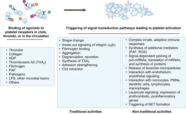
Platelets are essential for physiological hemostasis and are central in pathological thrombosis. These are their traditional and best known activities in health and disease. In addition, however, platelets have specializations that broaden their functional repertoire considerably. These functional capabilities, some of which are recently discovered, include the ability to sense and respond to infectious and immune signals and to act as inflammatory effector cells. Human platelets and platelets from mice and other experimental animals can link the innate and adaptive limbs of the immune system and act across the immune continuum, often also linking immune and hemostatic functions. Traditional and newly recognized facets of the biology of platelets are relevant to defensive, physiological immune responses of the lungs and to inflammatory lung diseases. The emerging view of platelets as blood cells that are much more diverse and versatile than previously thought further predicts that additional features of the biology of platelets and of megakaryocytes, the precursors of platelets, will be discovered and that some of these will also influence pulmonary immune defenses and inflammatory injury.


Figures

FIGURE 1.
FIGURE 1.
Platelets are effector cells in hemostasis, inflammation, and thrombotic and inflammatory syndromes in the lung and other organs. A: an organized thrombus occupying a medium size pulmonary artery in the lung of a patient who died of ARDS secondary to sepsis is shown. Neutrophils and monocytes are embedded in the thrombus. Primary hemostatic activities of platelets contribute to the formation of clots and thrombi, which are also sites of interaction with myeloid leukocytes and of other inflammatory and immune effector activities of platelets (see Figure 2). Pro-inflammatory responses of platelets, and platelet-leukocyte interactions, can be modeled in vitro (B and C). B: a platelet-fibrin complex formed by thrombin-stimulated human platelets incubated in the presence of extracellular fibrinogen in vitro. Interleukin-1β protein was detected in the activated platelets (yellow staining) and the fibrin mesh (orange staining) by immunocytochemistry. Green staining indicates actin. [From Lindemann et al. (249).] C: aggregates of thrombin-stimulated human platelets and isolated human monocytes that formed in vitro. The dark staining of the monocyte nuclei (arrows) indicates translocation of nuclear factor kappaB (NF-κB) from the leukocyte cytoplasm to the nucleus in response to molecular signals from the activated platelets. [From Weyrich et al. (464).] Signaling of monocytes by activated platelets induces expression of NF-κB-dependent chemokines and other inflammatory mediators by the leukocytes (453, 454, 464).
FIGURE 2.
FIGURE 2.
The functional repertoire of activated platelets includes multiple activities that mediate hemostasis and inflammation. In primary hemostasis, platelets are activated at sites of endothelial injury and exposure of subendothelial matrix. Platelets are also activated in evolving clots and thrombi and can be activated in the circulation in systemic thrombotic and inflammatory conditions. “Traditional” responses of activated platelets that are critical in physiological hemostasis and pathological thrombosis include shape change, inside-out signaling of integrin αIIbβ3 and other integrins, fibrinogen binding, aggregation, synthesis of thromboxane A2, degranulation and release of hemostatic mediators, adhesion strengthening, and clot retraction. Each of these traditional platelet hemostatic activities has documented or proposed inflammatory functions as well (see text and Table 1). Platelets also have additional biologic activities, many of which are relatively newly discovered, that can initiate or amplify inflammatory and immune responses. In many cases, these nontraditional biologic activities can also contribute to physiological or pathological hemostasis. Each response of activated platelets listed in this figure is discussed in the text. PAF, platelet activating factor; ROS, reactive oxygen species; IL-1β, interleukin-1β; PMNs, polymorphonuclear leukocytes; NET, neutrophil extracellular trap.
FIGURE 3.
FIGURE 3.
Activities of activated platelets may be critical in acute inflammatory lung injury in ARDS. Serial chest radiographs demonstrate increased permeability pulmonary edema that progressed over a 48-h interval from initial evaluation in the emergency department (A) until requirement for endotracheal intubation and mechanical ventilation (B) in a previously healthy patient with H1N1 influenza infection. Platelets may be key effector cells in increased alveolar capillary permeability (Figure 4) and alveolar inflammation (Figure 5) in ARDS caused by sterile and infectious triggers, including influenza. Experimental models of influenza indicate that platelets have nontraditional activities (Figure 2) in the lungs in this infection. See text for additional details. [From Matthay et al. (277).]
FIGURE 4.
FIGURE 4.
Platelets are critical in maintenance of endothelial barrier function but also induce increased endothelial permeability in inflammation. A: when platelet numbers are sufficient, semipermeable endothelial barriers that restrict transfer of water and proteins out of systemic and alveolar capillaries are maintained and protected. Release of stabilizing factors by platelets is one mechanism for endothelial barrier maintenance, although others have also been reported or proposed. Vascular endothelial cell cadherin (VE-cadherin) bonds are critical for establishing and maintaining alveolar and systemic endothelial barrier integrity. B: in severe thrombocytopenia, basal endothelial barrier properties are disrupted, leading to leak of water and protein from alveolar and systemic vessels. Large arrows indicate transvascular fluid and RBC escape into the alveolar space, and small arrows indicate leak into the interstitial and lymphatic space. The size of the arrows does not denote the relative magnitudes of the leaks into these compartments. Severe thrombocytopenia also contributes to inflammation-associated hemorrhage in experimental models. Platelet dysfunction, in addition to decreased platelet numbers, may have this effect. C: activated platelets can induce or amplify increased permeability of alveolar and systemic endothelial barriers in inflammation. Several mechanisms have been proposed or demonstrated in experimental models, including release of platelet factors that disrupt endothelial barriers, signaling of endothelial cells, and interaction with PMNs and monocytes, leading to disruption of endothelial bonds and leak of fluid, proteins, and RBC. Large and small arrows indicate escape of fluid and RBC as outlined in B. Increased permeability lung edema is a key feature of ARDS (Figure 3) and occurs in other syndromes of inflammatory lung injury. [Modified from Weyrich and Zimmerman (468).]
FIGURE 5.
FIGURE 5.
Platelets accumulate in the alveoli of mice in response to intrapulmonary LPS challenge. A: scattered platelets detected by brown immunostaining with an antibody against CD41 (αIIb subunit of integrin αIIbβ3) are present in alveolar vessels of control mice. (Scale bar = 20 μm; zoom of outlined area = original magnification ×60). Some large areas of anti-CD41 staining may indicate megakaryocytes. B: there was dramatic accumulation of CD41-positive platelets in alveoli of mice challenged with intratracheal LPS. Some CD41-positive staining may indicate platelet microparticles or megakaryocytes. In the right-hand enlarged panel, arrows point to CD41 events in the alveoli, many of which demonstrate platelets associated with intra-alveolar leukocytes. Platelets may have novel extravascular inflammatory activities in the alveolar space in addition to intravascular proinflammatory functions. [From Ortiz-Munoz et al. (330), with permission from American Society of Hematology.]
FIGURE 6.
FIGURE 6.
Activated human platelets synthesize IL-1β in response to hemostatic and inflammatory agonists and dengue virus. Human platelets synthesize IL-1β when activated by hemostatic and inflammatory agonists including thrombin, collagen, ADP, PAF, and LPS. In addition, live dengue virus induces synthesis of IL-1β by engaging DC-SIGN. The mechanism of IL-1β synthesis by activated human platelets involves signal-dependent, spliceosome-mediated splicing of the IL-1β pre-mRNA transcript followed by signal-dependent translation of the mature mRNA and generation of pro-IL-1β protein. Newly synthesized pro-IL-1β is processed to IL-1β by NLRP3 inflammasome-dependent caspase-1 activity, as shown in DENV-activated platelets and platelets from DENV-infected patients. Mature, biologically active IL-1β protein is released from activated platelets in solution and in microparticles. IL-1β in microparticles from activated platelets induces increased permeability of human endothelial monolayers and other inflammatory responses of human endothelial cells. Increased vascular permeability in patients with DENV infection correlated with platelet and platelet microparticle IL-1β and with platelet caspase-1 activity. See text for details and references. [Adapted from Hottz et al. (179).]
FIGURE 7.
FIGURE 7.
Activated platelets interact with endothelial cells and leukocytes, providing mechanisms for intercellular signaling and induction and amplification of effector responses in inflammation. A: activation of platelets induces platelet-platelet aggregation (see Figure 1B) and intercellular signaling. Platelet aggregation occurs in hemostasis, thrombosis, and inflammation. Platelets and platelet aggregates adhere to and signal endothelial cells in these conditions based on experimental observations. Activated platelets also adhere to leukocytes, forming mixed cell aggregates (“heterotypic aggregates”) that can be retrieved from the blood and detected in vessels and at extravascular sites by histology and intravital microscopy. Platelet-leukocyte aggregates are vehicles for complex signaling and synthesis of inflammatory and hemostatic mediators (see Figure 1C). Microvessels in the lung are sites of platelet-leukocyte aggregate formation and accumulation of platelets and platelet-leukocyte aggregates based on intravital microscopic observations in experimental animals (see FIGURE 5). This may also be an important mechanism in clinical acute lung injury and ARDS. Platelets also interact with leukocytes in clots and thrombi (see Figure 1A). B: activated platelets signal and form aggregates with multiple leukocyte types, including neutrophils (PMNs), eosinophils, monocytes, and lymphocyte subclasses. This is one of several mechanisms that enable platelets to act across the immune continuum, mediate events in the innate and adaptive limbs of the immune system, and link hemostasis and inflammation. C: activated platelets also interact with macrophages (not shown) and dendritic cells, which have multiple regulatory activities in innate and adaptive immune function. Platelets influence dendritic cell differentiation, maturation, and interaction with T- and B-lymphocytes and interact with dendritic cell subsets in the blood. [B and C from Vieira-de-Abreu et al. (454), with permission from Springer Science + Business Media.]
FIGURE 8.
FIGURE 8.
Activated platelets trigger NET formation by neutrophils. Pathogens (bacteria, viruses, fungi, others), chemokines, and other host mediators as well as activated platelets trigger NETosis and NET formation in experimental models. Experimental, and limited clinical, observations indicate that NET formation is a mechanism of extracellular capture, containment, and potential killing of pathogens in vessels and alveoli of the lungs, and in other organs. In addition, however, NET formation can mediate vascular and acute lung injury based on experimental studies. NET-associated factors including histones, neutrophil elastase, and other granule enzymes injure endothelial and alveolar epithelial cells in vitro and in vivo. NETs also induce thrombosis and are components of clots. NET formation may contribute to ARDS and other syndromes of inflammatory injury to the lungs as well as to venous thromboembolism.
FIGURE 9.
FIGURE 9.
Platelets have sentinel and effector capabilities that allow them to sense and respond to pathogens and microbial products. Platelets utilize a variety of receptors and signal transduction pathways in responses to bacteria, viruses, and other pathogens and have a diverse array of antimicrobial defensive mechanisms. Pathogen-induced platelet activation can also injure the host. Human platelets incubated alone, with Escherichia coli, or with Staphylococcus aureus for 30 min and stained for F-actin and bacterial DNA are shown in this figure. Live E. coli and S. aureus, E. coli LPS, and α-toxin from S. aureus induce a variety of functional responses by platelets in vitro, including signal-dependent protein synthesis (Figure 2). [From Rondina et al. (367).]

References

    1. Aggrey AA, Srivastava K, Ture S, Field DJ, Morrell CN. Platelet induction of the acute-phase response is protective in murine experimental cerebral malaria. J Immunol : 4685–4691, 2013. - PMC - PubMed
    1. Alexander JS, Patton WF, Christman BW, Cuiper LL, Haselton FR. Platelet-derived lysophosphatidic acid decreases endothelial permeability in vitro. Am J Physiol Heart Circ Physiol : H115–H122, 1998. - PubMed
    1. Alitalo K. The lymphatic vasculature in disease. Nature Med : 1371–1380, 2011. - PubMed
    1. Ammon C, Kreutz M, Rehli M, Krause SW, Andreesen R. Platelets induce monocyte differentiation in serum-free coculture. J Leukocyte Biol : 469–476, 1998. - PubMed
    1. Anabel AS, Eduardo PC, Pedro Antonio HC, Carlos SM, Juana NM, Honorio TA, Nicolas VS, Sergio Roberto AR. Human platelets express Toll-like receptor 3 and respond to poly I:C. Hum Immunol : 1244–1251, 2014. - PubMed

Publication types