. 2016 Jun 30:14:e0184.
doi: 10.1199/tab.0184. eCollection 2016.

Biotrophy at Its Best: Novel Findings and Unsolved Mysteries of the Arabidopsis-Powdery Mildew Pathosystem

Affiliations

Biotrophy at Its Best: Novel Findings and Unsolved Mysteries of the Arabidopsis-Powdery Mildew Pathosystem

Hannah Kuhn et al. Arabidopsis Book. .

Abstract

It is generally accepted in plant-microbe interactions research that disease is the exception rather than a common outcome of pathogen attack. However, in nature, plants with symptoms that signify colonization by obligate biotrophic powdery mildew fungi are omnipresent. The pervasiveness of the disease and the fact that many economically important plants are prone to infection by powdery mildew fungi drives research on this interaction. The competence of powdery mildew fungi to establish and maintain true biotrophic relationships renders the interaction a paramount example of a pathogenic plant-microbe biotrophy. However, molecular details underlying the interaction are in many respects still a mystery. Since its introduction in 1990, the Arabidopsis-powdery mildew pathosystem has become a popular model to study molecular processes governing powdery mildew infection. Due to the many advantages that the host Arabidopsis offers in terms of molecular and genetic tools this pathosystem has great capacity to answer some of the questions of how biotrophic pathogens overcome plant defense and establish a persistent interaction that nourishes the invader while in parallel maintaining viability of the plant host.


Figures
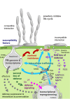
Figure 1.
Figure 1.
Graphical overview of the topics discussed in this article. EHM, extrahaustorial membrane; EHMx, extrahaustorial matrix; ER, endoplasmic reticulum; JA, jasmonic acid; MAPK(KK/KKK), mitogen-activated protein kinase (kinase kinase/kinase kinase kinase); MVBs, multivesicular bodies; PRRs, pattern recognition receptors; RPW8, RESISTANCE TO POWDERY MILDEW 8 protein; ROS, reactive oxygen species; SA, salicylic acid; TGN, trans-Golgi network. See text for further explanations.
Figure 2.
Figure 2.
Asexual life cycle of G. orontii in association with Arabidopsis. The central part of the figure illustrates schematically the key steps of the life cycle, while the micrographs show the actual fungal infection structures. The confocal laser scanning micrographs were obtained from transgenic Col-0 plants stably expressing yellow cameleon inoculated with Go. Fungal infection structures were stained with FM4-64 (shown in red) while green fluorescence is representative of cytosolic yellow cameleon fluorescence. Bars: 20 µm.
Figure 3.
Figure 3.
The PM haustorium. The fungal haustorium forms within cells of the leaf epidermis after penetration. A. Scheme of a PM haustorium (grey) separated from the plant cytoplasm by fungal haustorial membrane (fHM), fungal cell wall (fCW), extrahaustorial matrix (EHMx) and extrahaustorial membrane (EHM). The inset depicts the proposed exocytosis of fungal multivesicular bodies (fMVBs) B. Wheat germ agglutinin staining of chitin in an isolated haustorium of Go. The confocal laser scanning micrograph shows a mature haustorium body (HB) with numerous haustorial lobes (L). C. Partial callose encasement of an isolated Go haustorium. The electron-opaque EHM (arrowheads) surrounds the haustorium (H) but not the callose-containing encasement (E). Bars: B 5 µm; C 2 µm. Panels B and C reproduced with permission from (Micali et al., 2011) (Copyright by John Wiley & Sons (Cellular Microbiology)).
Figure 4.
Figure 4.
Nanoscale resolution of callose polymer fibrils in pathogen-induced cell wall papillae. Three-week-old Arabidopsis wild type and pathogen-resistant PMR4-GFP overexpressing lines (P35S::PMR4-GFP) were inoculated with the adapted powdery mildew Gc. Localization microscopy (dSTORM: direct stochastical optical reconstruction microscopy) of aniline blue-stained callose polymer fibrils in pathogen-induced papillae at sites of attempted fungal penetration at 12 hpi in rosette leaves. Scale bars = 2 µm. Unpublished micrograph, courtesy of Christian A. Voigt and Dennis Eggert.
Figure 5.
Figure 5.
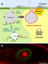
RPW8 localizes at the EHM and contributes to cell death upon PM infection. A. Scheme depicting RPW8.2 function. Left: RPW8.2 interactors and RPW8.2 deposition at the EHM. Right: RPW8.2-triggered oxidative burst and callose-encasement of haustoria correlates with subsequent host cell death. B. Confocal laser scanning micrograph of GFP-labelled RPW8.2 (green) in the EHM. Red, propidium iodide stained plant and fungal structures. Bar = 10 µm. Unpublished micrograph, courtesy of Wenming Wang and Shunyuan Xiao.
Figure 6.
Figure 6.
Macroscopic infection phenotypes of Col-0 and the mlo2 mlo6 mlo12 mutant. Five-week-old wild type (Col-0) and mlo2 mlo6 mlo12 plants (in Col-0 genetic background) were inoculated with Go and photographs were taken one week after inoculation.
Figure 7.
Figure 7.
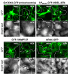
Myosin inhibition affects the recruitment of organelles and endo-membrane compartments to PM attack sites. Leaves stably expressing GFP fusions of (i) Saccharomyces cerevisiae cytochrome c oxidase IV (Pd35S::ScCOX4-GFP; a mitochondrial marker; Nelson et al., 2007), (ii) the signal peptide of WALL-ASSOCIATED KINASE 2 together with the ER retention signal HDEL (Pd35S::SPAtWAK2-GFP-HDEL; an ER marker; Nelson et al., 2007), (iii) the Rab5 GTPase ARA6/RAB1F (PARA6::ARA6-GFP; an endomembrane vesicle marker; Goh et al., 2007), and (iv) the v-SNARE VAMP727 (PVAMP727::GFP-VAMP727; an endomembrane vesicle marker; Ebine et al., 2008) were infiltrated with water (mock) or 1 mM N-ethylmaleimide (NEM; a myosin inhibitor) and 1 h later inoculated with Bgh. Infected epidermal cells (indicated by dashed lines) were examined by confocal microscopy ca. 16 hpi. Projections of z-stacks are shown. Asterisks indicate the Bgh penetration site. Bar = 10 µm. Unpublished micrographs, courtesy of Yangdou Wei.
Figure 8.
Figure 8.
Integration of phytohormone signaling in defense against PM fungi. Although only incompatible PM-host interactions elicit JA/ET-mediated defense, JA/ET-induced defense responses are effective against virulent PM fungi if stimulated constitutively (cesA/cev1), artificially (JA treatment) or systemically (Piriformospora indica root colonization). These findings suggest that virulent fungi suppress JA/ET signaling during compatible interactions. This suppression might involve the antagonistic action of SA signaling. Solid lines indicate experimentally supported impacts, while dashed lines indicate speculative connections.
Figure 9.
Figure 9.
Proposed model of endoreduplication in PM pathogenesis. The scheme depicts regulators and mechanisms involved in the control of endoreduplication and consequences of PM-induced mesophyll polyploidy. A PM (grey)-colonized leaf epidermal cell and an underlying mesophyll cell are shown. Grey arrows indicate the proposed translocation of components between cells. Solid lines indicate proven regulatory impacts and dashed lines indicate speculative regulatory impacts. M = mitosis.

References

    1. Acevedo-Garcia J., Kusch S., and Panstruga R. ( . 2014). Magical mystery tour: MLO proteins in plant immunity and beyond. New Phytol. 204: 273– 281. - PubMed
    1. Adam L., and Somerville S.C. ( . 1996). Genetic characterization of five powdery mildew disease resistance loci in Arabidopsis thaliana. Plant J. 9: 341– 356. - PubMed
    1. Adam L., Ellwood S., Wilson I., Saenz G., Xiao S., Oliver R.P., Turner J.G., and Somerville S. ( . 1999). Comparison of Erysiphe cichoracearum and E. cruciferarum and a Survey of 360 Arabidopsis thaliana Accessions for Resistance to These Two Powdery Mildew Pathogens. Mol. Plant Microbe Interact. 12: 1031– 1043. - PubMed
    1. An Q., Hückelhoven R., Kogel K.H., and van Bel A.J. ( . 2006). Multivesicular bodies participate in a cell wall-associated defence response in barley leaves attacked by the pathogenic powdery mildew fungus. Cell Microbiol. 8: 1009– 1019. - PubMed
    1. Andersson M.X., Kourtchenko O., Dangl J.L., Mackey D., and Ellerström M. ( . 2006). Phospholipase-dependent signalling during the AvrRpm1-and AvrRpt2-induced disease resistance responses in Arabidopsis thaliana. Plant J. 47: 947– 959. - PubMed

LinkOut - more resources