. 2016 Aug 2:7:16.
doi: 10.1186/s13227-016-0054-6. eCollection 2016.

The developmental and genetic bases of apetaly in Bocconia frutescens (Chelidonieae: Papaveraceae)

Affiliations

The developmental and genetic bases of apetaly in Bocconia frutescens (Chelidonieae: Papaveraceae)

Cristina Arango-Ocampo et al. Evodevo. .

Abstract

Background: Bocconia and Macleaya are the only genera of the poppy family (Papaveraceae) lacking petals; however, the developmental and genetic processes underlying such evolutionary shift have not yet been studied.

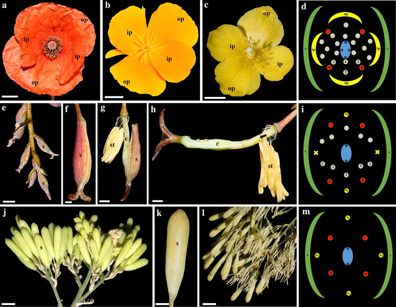
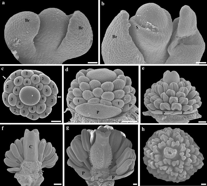
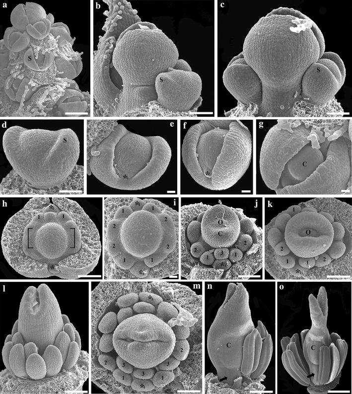
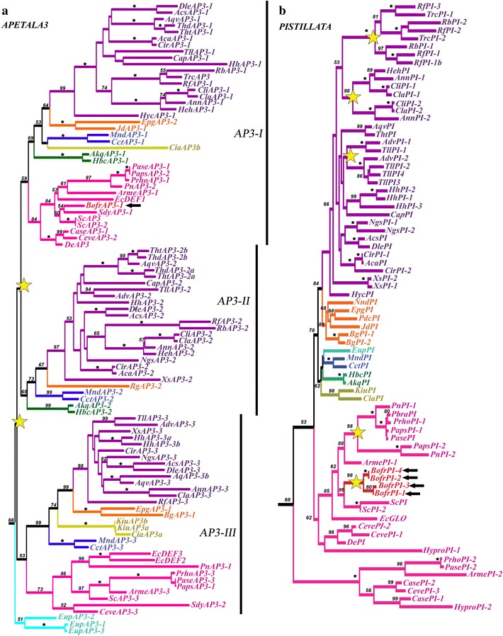
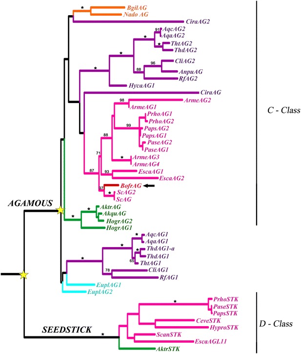
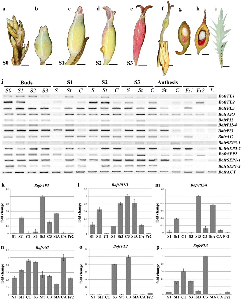
Results: We studied floral development in two species of petal-less poppies Bocconia frutescens and Macleaya cordata as well as in the closely related petal-bearing Stylophorum diphyllum. We generated a floral transcriptome of B. frutescens to identify MADS-box ABCE floral organ identity genes expressed during early floral development. We performed phylogenetic analyses of these genes across Ranunculales as well as RT-PCR and qRT-PCR to assess loci-specific expression patterns. We found that petal-to-stamen homeosis in petal-less poppies occurs through distinct developmental pathways. Transcriptomic analyses of B. frutescens floral buds showed that homologs of all MADS-box genes are expressed except for the APETALA3-3 ortholog. Species-specific duplications of other ABCE genes in B. frutescens have resulted in functional copies with expanded expression patterns than those predicted by the model.

Conclusions: Petal loss in B. frutescens is likely associated with the lack of expression of AP3-3 and an expanded expression of AGAMOUS. The genetic basis of petal identity is conserved in Ranunculaceae and Papaveraceae although they have different number of AP3 paralogs and exhibit dissimilar floral groundplans.

Keywords: ABCE model; AGAMOUS; APETALA3; Apetaly; Bocconia; Homeosis; Macleaya; Papaveraceae; Stylophorum.


Figures

Fig. 1
Fig. 1
Floral diversity in Papaveraceae. Petal-bearing flowers in Papaver rhoeas (a), Eschscholzia californica (b) and Stylophorum diphyllum (c). d S. diphyllum floral diagram. em Petal-less flowers. eh Bocconia frutescens, preanthetic and anthetic flowers. i B. frutescens floral diagram. jl Macleaya cordata, preanthetic and anthetic flowers. m M. cordata floral diagram. Arrow gynophore; c carpel, ip inner petal, op outer petal, s sepal, st stamen, * homeotic stamen; 1, first set of 4 true stamens on the first whorl in red indicate that these are the first stamens formed and that they occupy the same positions in both petalous and apetalous species serving as reference for the positioning of all other stamens; 2, second set of true stamens of the first whorl; 3, true stamens of the second whorl. 4. True stamens of the third whorl. Bars ac = 7 mm; e, g, h = 10 mm; f = 2 mm, j, l = 8 mm; k = 4 mm
Fig. 2
Fig. 2
Scanning electron micrographs of floral development of Stylophorum diphyllum. a Flower primordium. b Sepal initiation. c, d Initiation of petals (arrows in c), stamens (numbered) and gynoecium. eh Successive growth stages of stamens and gynoecium; some stamens removed in f and g. Br bract, C carpel, P petal, S sepal; 1, 2, first and second sets of the first whorl; 3, stamens of the second whorl; 4, stamens of the third whorl. Bars ac = 50 µm; dh = 100 µm
Fig. 3
Fig. 3
Scanning electron micrographs of floral development of Bocconia frutescens. a Developing inflorescence. b, c Detail of the apex of the inflorescence; note the larger terminal flower with respect to the two lateral flowers. d Initiation of the sepals. e, f Initiation of the two homeotic stamens (*1). h Initiation of the first whorl of true stamens alternate to the sepals (1); note the two opposite common primordia ([ ]). i Initiation of the second (inner) set of true stamens of the first whorl, developed from common primordial. jm Initiation of the third set of true stamens belonging to the second whorl (3) and the ovule. n, o Late development of stamens and gynoecium; note the conspicuous stigmatic tissue, the horse-shoe-shaped valve on each carpel and the gynophore (arrow); C carpel, O ovule, S sepal, * homeotic stamens; 1, first whorl of true stamens; 2, second set of true stamens belonging to the first whorl; 3, third set of true stamens belonging to the second whorl. Bars ad, h, jo = 50 µm; eg, i = 10 µm
Fig. 4
Fig. 4
Scanning electron micrographs of floral development of Macleaya cordata. a Developing inflorescence; note the larger terminal flower. b Initiation of sepals. c, d Initiation of the first whorl of homeotic stamens, alternate to the sepals (*1). eg Successive initiation of the second whorl of homeotic stamens, opposite to the sepals (*2), and the single whorl of four, decussate true stamens (1). hj Growth of the stamens and the short, sessile gynoecium. C Carpel, S sepal, * homeotic stamens; 1, first whorl of true stamens. Bars a, j = 100 µm; bg = 10 µm; h, i = 50 µm
Fig. 5
Fig. 5
Maximum likelihood trees of APETALA3 (a) and PISTILLATA (b) in Ranunculales. Colors indicate the following taxa: light blue Eupteleaceae; pink Papaveraceae; green Lardizabalaceae; straw Circaeasteraceae; dark blue Menispermaceae; orange Berberidaceae; purple Ranunculaceae. Stars indicate gene duplications. Arrows point to the single copy of AP3 (BofrAP3-1 in a) and the four PI paralogs (BofrPI1/BofrPI2/BofrPI3/BofrPI4 in b) in B. frutescens. Bootstrap values (BS) above 50 are placed at nodes; asterisks indicate BS of 100
Fig. 6
Fig. 6
Maximum likelihood trees of FUL-like (a) and SEPALLATA (b) in Ranunculales. Colors indicate the following taxa: light blue Eupteleaceae; pink Papaveraceae; green Lardizabalaceae; dark blue Menispermaceae; orange Berberidaceae; purple Ranunculaceae. Stars indicate gene duplications. Arrows point to three FUL-like paralogs (BofrFL1/BofrFL2/BofrFL3 in a) and five SEP copies (BofrSEP1-1, BofrSEP1-2, BofrSEP2, BofrSEP3-1 and BofrSEP3-2 in b) in B. frutescens. Bootstrap values (BS) above 50 are placed at nodes; asterisks indicate BS of 100
Fig. 7
Fig. 7
Maximum likelihood tree of AGAMOUS in Ranunculales. Colors indicate the following taxa: light blue Eupteleaceae; pink Papaveraceae; green Lardizabalaceae; dark blue Menispermaceae; orange Berberidaceae; purple Ranunculaceae. Stars indicate gene duplications. Arrow points to the single copy of AG (BofrAG) in B. frutescens. Bootstrap values (BS) above 50 are placed at nodes; asterisks indicate BS of 100
Fig. 8
Fig. 8
Floral stages and dissected organs of B. frutescens with corresponding expression studies. Inflorescence (a) and floral bud (b) at Stage 0 (S0). Floral buds at stages S1 (c), S2 (d) and S3 (e). f Flowers at anthesis. Young (Fr1) (g) and mature fruits (Fr2) (h). i Leaves. j Locus-specific RT-PCR results for all isolated MADS-box genes. Sepals (S), stamens (St) and carpels (C) were also dissected individually at stages S1, S2, S3. ACTIN (BofrACT) was used as a positive control. L Leaf. Numbers after S and Fr correspond to the different developmental stages. Relative expression patterns of BofrAP3 (k), BofrPI1/3 (l), BofrPI2/4 (m), BofrAG (n), BofrFL2 (o), BofrFL3 (p) in selected floral organs. Values are mean ± SD for three technical replicates. Bars in ai = 0.5 mm
Fig. 9
Fig. 9
Representative scheme of the A, B, C and E-class gene expression in petalous (Eschscholzia californica and Papaver somniferum) and apetalous (Bocconia frutescens) Papaveraceae members in comparison with the model Ranunculaceae (Aquilegia coerulea). Light yellow in Aquilegia for homologs of FUL-like genes indicates broad expression in floral organs but no known roles in floral organ identity [27]. The models are taken directly or constructed based on data from Drea et al. [28], Yellina et al. [54], Pabón-Mora et al. [26, 27], Sharma and Kramer [29], Lange et al. [55]. St.odia Staminodia

References

    1. Endress PK. Diversity and evolutionary biology of tropical flowers. Cambridge: Cambridge University Press; 1994.
    1. Endress PK. Floral structure and evolution in Ranunculanae. Plant Syst Evol. 1995;9:47–61.
    1. Ronse De Craene LP. Are petals sterile stamens or bracts? The origin and evolution of petals in the core eudicots. Ann Bot. 2007;100:621–630. doi: 10.1093/aob/mcm076. - DOI - PMC - PubMed
    1. Ronse De Craene LP, Brockington SF. Origin and evolution of petals in angiosperms. Plant Ecol Evol. 2013;146:5–25. doi: 10.5091/plecevo.2013.738. - DOI
    1. Glover BJ. Understanding flowers and flowering: an integrated approach. New York: Oxford University Press; 2007.

LinkOut - more resources