. 2016 Aug 2:6:48.
doi: 10.1186/s13578-016-0115-5. eCollection 2016.

EphA5 and EphA6: regulation of neuronal and spine morphology

Affiliations

EphA5 and EphA6: regulation of neuronal and spine morphology

Gitanjali Das et al. Cell Biosci. .

Abstract

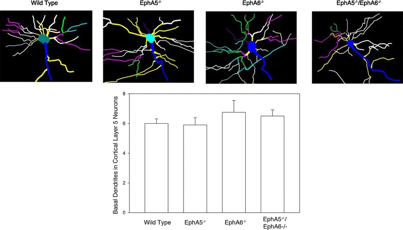
Background: The Eph family of receptor tyrosine kinases plays important roles in neural development. Previous studies have implicated Eph receptors and their ligands, the ephrins, in neuronal migration, axon bundling and guidance to specific targets, dendritic spine formation and neural plasticity. However, specific contributions of EphA5 and EphA6 receptors to the regulation of neuronal cell morphology have not been well studied.

Results: Here we show that deletion of EphA5 and EphA6 results in abnormal Golgi staining patterns of cells in the brain, and abnormal spine morphology.

Conclusion: These observations suggest novel functions of these Eph receptors in the regulation of neuronal and spine structure in brain development and function.

Keywords: Cortex; Dendrite; EphA5; EphA6; Golgi staining; Spine.


Figures

Fig. 1
Fig. 1
Representative images of the beta-galactosidase staining in different regions of the mouse brain showing the expression of EphA5 (ac, g, h) and EphA6 (df, i, j) genes. Highest expression of both genes is present in the cortical regions with also diffuse presence in the thalamus, hypothalamus, hippocampus and amygdala as well (n = 3 for both EphA5 and EphA6 brains). Amyg amygdala; CP Caudate Putamen; CTX cerebral cortex; HIP hippocampus; HY hypothalamus; TH thalamus; s septum; OLF olfactory area; PIR pyriform cortex; Scale bars af, 1 mm; g, i, 0.5 mm: h, j, 1 mm
Fig. 2
Fig. 2
Representative images of Golgi staining done in paraformaldehyde perfused brains showing aggregation of neurons in the frontal cortical (upper panels) and mid-cortical (lower panels) regions of both EphA5 and EphA6 KO brains. The double knockout (DKO) of EphA5 and EphA6 did not show a more pronounced effect on this aggregation phenomenon. The bracket areas show approximate locations of the cortex that are examined in higher magnification in Fig. 3. Scale bars 1 mm
Fig. 3
Fig. 3
Representative higher magnification images of Golgi staining done in paraformaldehyde perfused brain showing aggregation of neurons at the frontal cortical (upper panels) and the mid-cortical (lower panels) regions of both EphA5 and EphA6 KO brains. The double knockout (DKO) of EphA5 and EphA6 did not show a more pronounced effect on this aggregation phenomenon. Scale bar 500 µm
Fig. 4
Fig. 4
Representative images of Golgi staining done in fresh non-paraformaldehyde perfused brains showing aggregation of neurons in the frontal cortical (upper panels) and the mid-cortical (lower panels) regions of both EphA5 and EphA6 KO brains. The double knockout (DKO) of EphA5 and EphA6 did not show a more pronounced effect on this aggregation phenomenon. Golgi staining of the fresh brains clearly showed that the aggregation phenomenon is mostly a neuronal effect. The bracket areas show approximate locations of the cortex that are examined in higher magnification in Fig. 5. Scale bars 1 mm
Fig. 5
Fig. 5
Representative higher magnification images of Golgi staining done in fresh non- paraformaldehyde perfused brains showing aggregation of neurons at the frontal cortical (upper panels) and the mid-cortical (lower panels) regions of both EphA5 and EphA6 KO brains. The double knockout (DKO) of EphA5 and EphA6 did not show a more pronounced effect on this aggregation phenomenon. Golgi staining of the fresh brains clearly showed that the aggregation phenomenon is mostly a neuronal effect. Scale bar 500 µm
Fig. 6
Fig. 6
Double immunostaining showed the presence of neuronal clumps (NeuN in green) in the cortical layer 5 in all the knockout types. Most of the clumps were neuronal in origin with a few NeuN-negative cells as well (yellow arrows). a wild type; b EphA5−/−; c EphA6−/−; d EphA5−/−EphA6−/−. Scale bars 20 µm
Fig. 7
Fig. 7
Representative neurolucida drawings of the basal dendrite of the different genotypes in the cortical layer 5 (upper panel). Statistical analysis showed no difference in the number of basal dendrites amongst different genotypes (lower panel). Total of 60 neurons of each genotype was used for the analysis (three animals, 20 neurons each)
Fig. 8
Fig. 8
Representative images of spines in the basal dendrites of neurons in cortical layer 5. Wild type spines showed the presence of stubby, filamentous as well as mushroom type of spines, while the knockouts, displaying an irregular morphology, are difficult to classify into the classic categories. Scale bar 20 µm

References

    1. Pitulescu ME, Adams RH. Eph/ephrin molecules—a hub for signaling and endocytosis. Genes Dev. 2010;24(22):2480–2492. doi: 10.1101/gad.1973910. - DOI - PMC - PubMed
    1. Flanagan JG, Vanderhaeghen P. The ephrins and Eph receptors in neural development. Ann Rev Neurosci. 1998;21:309–345. doi: 10.1146/annurev.neuro.21.1.309. - DOI - PubMed
    1. Kullander K, Klein R. Mechanisms and functions of Eph and ephrin signalling. Nat Rev Mol Cell Biol. 2002;3(7):475–486. doi: 10.1038/nrm856. - DOI - PubMed
    1. O’Leary DD, Wilkinson DG. Eph receptors and ephrins in neural development. Curr Opin Neurobiol. 1999;9(1):65–73. doi: 10.1016/S0959-4388(99)80008-7. - DOI - PubMed
    1. Pasquale EB. Eph receptor signalling casts a wide net on cell behaviour. Nat Rev Mol Cell Biol. 2005;6(6):462–475. doi: 10.1038/nrm1662. - DOI - PubMed

LinkOut - more resources