. 2016 Jul 18:3:145-52.
doi: 10.1016/j.ejro.2016.06.003. eCollection 2016.

Locally advanced rectal cancer: Qualitative and quantitative evaluation of diffusion-weighted MR imaging in the response assessment after neoadjuvant chemo-radiotherapy

Affiliations

Locally advanced rectal cancer: Qualitative and quantitative evaluation of diffusion-weighted MR imaging in the response assessment after neoadjuvant chemo-radiotherapy

Pietro Valerio Foti et al. Eur J Radiol Open. .

Abstract

Purpose: to investigate the added value of qualitative and quantitative evaluation of diffusion weighted (DW) magnetic resonance (MR) imaging in response assessment after neoadjuvant chemo-radiotherapy (CRT) in patients with locally advanced rectal cancer (LARC).

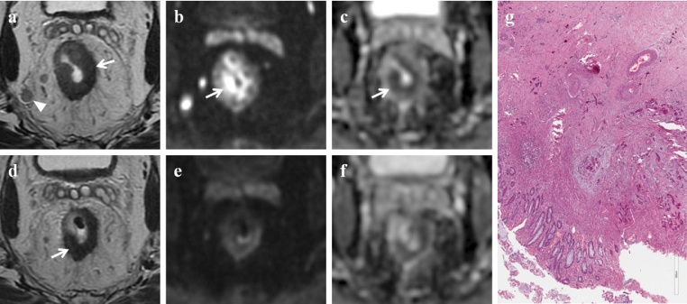
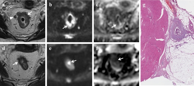
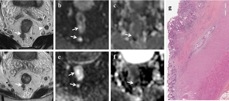
Methods: 31 patients with LARC (stage ≥ T3) were enrolled in the study. All patients underwent conventional MRI and DWI before starting therapy and after neoadjuvant CRT. All patients underwent surgery; pathologic staging represented the reference standard. For qualitative analysis, two radiologists retrospectively reviewed conventional MR images and the combined set of conventional and DW MR images and recorded their confidence level with respect to complete response (ypCR). For quantitative analysis, tumor's apparent diffusion coefficient (ADC) values were measured at each examination. ADC pre-CRT, ADC post-CRT and Δ ADC post-ADC pre of the three groups of response (ypCR, partial response ypPR, stable disease ypSD) were compared. Receiver-operating characteristics (ROC) curve analysis was employed to investigate the discriminatory capability for ypCR, responders (ypCR, ypPR) and ypSD of each measure.

Results: addition of DWI to conventional T2-weighted sequences improved diagnostic performance of MRI in the evaluation of ypCR. A low tumor ADC value in the pre-CRT examination, a high ADC value in the post-CRT examination, a high Δ ADC post-ADC pre [>0.3 (×10(-3) mm(2)/s)] were predictive of ypCR.

Conclusions: DW sequences improve MR capability to evaluate tumor response to CRT. Nevertheless, no functional MR technique alone seems accurate enough to safely select patients with ypCR.

Keywords: Chemoradiation; Diffusion-weighted imaging; Magnetic resonance imaging; Rectal cancer; Staging; Treatment response.


Figures

Fig. 1
Fig. 1
Radiotherapy plan. Radiotherapy dose distribution in axial, coronal and sagittal views. Planning was made through a 3D-conformal technique.
Fig. 2
Fig. 2
Complete response. MR images of a 70-year old man classified as complete responder on combined set of conventional and DW images and as partial responder on T2-weighted images. (a) Pre-CRT axial T2-weighted image shows neoplastic tissue in the middle rectum (white arrow) with nodal involvement (white arrowhead); the tumor has spread through the rectal wall into the perirectal fat for less than 5 mm (T3a N2 stage). (b) Pre-CRT DW (b = 800 s/mm2) image shows a focal high signal intensity area in the corresponding tumor (white arrow). (c) Pre-CRT ADC map at the same level shows reciprocal low signal intensity area due to the pathological tissue (white arrow). The mean ADC value was 0.752 × 10−3 s/mm2. (d) Post-CRT axial T2-weighted image shows a wall thickening of the rectum with low signal intensity (white arrow), not clearly depicted as fibrosis. (e) Post-CRT DW (b = 800 s/mm2) image shows no residual high signal intensity in the primary tumor bed. (f) Post-CRT ADC map at the same level: the mean ADC value was 1.076 × 10−3 s/mm2. On the basis of qualitative and quantitative DWI analysis the patient should be considered as complete responder. (g) Photomicrograph (H&E x800). Complete response (TRG: 4), absence of tumor remnants; fibrous reaction induced by the treatment and calcifications are evident.
Fig. 3
Fig. 3
Partial response. MR images of a 55-year old woman classified as partial responder both on combined set of conventional and DW images and on T2-weighted images. (a) Pre-CRT axial T2-weighted image shows wall thickening of the middle rectum (white arrow) with spread into the perirectal fat for less than 10 mm; there are also some lymph nodes (white arrowhead) into the mesorectal fat (T3b N2). (b) Pre-CRT DW (b = 800 s/mm2) image shows high signal intensity area in the corresponding tumor (white arrow). (c) Pre-CRT ADC map at the same level shows low signal intensity area due to the pathological tissue (white arrow). The mean ADC value was 0.804 × 10−3 s/mm2. (d) Post-CRT axial T2-weighted image shows tumor shrinkage and intermediate signal intensity tissue in the rectal wall, not clearly depicted as fibrosis. (e) Post-CRT DW (b = 800 s/mm2) image shows reduction of the high signal intensity area, which is now limited to a focal spot in the rectal wall (white arrow). (f) Post-CRT ADC map at the same level shows a focal low signal intensity area corresponding to the residual tumor (white arrow). The mean ADC value was 1.237 × 10−3 s/mm2. (g) Photomicrograph (H&E x800). Intermediate regression (TRG: 2 + 3), predominance of the fibrous reaction induced by the treatment and few tumor remnants.
Fig. 4
Fig. 4
Stable disease. MR images of a 77-year old man classified as stable disease both on combined set of conventional and DW images and on T2-weighted images. (a) Pre-CRT axial T2-weighted image shows neoplastic tissue in the middle rectum (white arrow) with nodal involvement (white arrowhead). The tumor has spread into the mesorectal fat for more than 10 mm (T3c N1 stage). (b) Pre-CRT DW (b = 800 s/mm2) image shows high signal intensity area due to neoplastic tissue (white arrows). (c) Pre-CRT ADC map at the same level shows low signal intensity area (white arrow). The mean ADC value was 0.937 × 10−3 s/mm2. (d) Post-CRT axial T2-weighted image shows a poor tumor reduction with persistence of neoplastic spread (white arrow) into the mesorectal fat (T3 stage). (e) Post-CRT DW (b = 800 s/mm2) image shows high signal intensity area in the corresponding tumor (white arrows). (f) Post-CRT ADC map at the same level shows low signal intensity area (white arrow). (g) Photomicrograph (H&E x600). Poor regression (TRG: 0 + 1), predominance of areas with tumor remnants surrounded by a poor fibrous reaction induced by the treatment.

References

    1. Maier A., Fuchsjäger M. Preoperative staging of rectal cancer. Eur. J. Radiol. 2003;47:89–97. - PubMed
    1. Attenberger U.I., Pilz L.R., Morelli J.N., Hausmann D., Doyon F., Hofheinz R., Kienle P., Post S., Michaely H.J., Schoenberg S.O., Dinter D.J. Multi-parametric MRI of rectal cancer—do quantitative functional MR measurements correlate with radiologic and pathologic tumor stages? Eur. J. Radiol. 2014;83:1036–1043. - PubMed
    1. Maas M., Nelemans P.J., Valentini V. Long-term outcome in patients with a pathological complete response after chemoradiation for rectal cancer: a pooled analysis of individual patient data. Lancet Oncol. 2010;11:835–844. - PubMed
    1. Semedo L.C., Lambregts D.M., Maas M. Rectal cancer: assessment of complete response to preoperative combined radiation therapy with chemotherapy conventional MR volumetry versus diffusion-weighted MR imaging. Radiology. 2011;260:734–743. - PubMed
    1. Kluza E., Rozeboom E.D., Maas M., Martens M., Lambregts D.M., Slenter J., Beets G.L., Beets-Tan R.G. T2 weighted signal intensity evolution may predict pathological complete response after treatment for rectal cancer. Eur. Radiol. 2013;23:253–261. - PubMed

LinkOut - more resources