

Fiber tracking: A qualitative and quantitative comparison between four different software tools on the reconstruction of major white matter tracts
- PMID: 27489869
- PMCID: PMC4959946
- DOI: 10.1016/j.ejro.2016.06.002
Fiber tracking: A qualitative and quantitative comparison between four different software tools on the reconstruction of major white matter tracts
Abstract
Purpose: Diffusion tensor imaging (DTI) enables in vivo reconstruction of white matter (WM) pathways. Considering the emergence of numerous models and fiber tracking techniques, we herein aimed to compare, both quantitatively and qualitatively, the fiber tracking results of four DTI software (Brainance, Philips FiberTrak, DSI Studio, NordicICE) on the reconstruction of representative WM tracts.
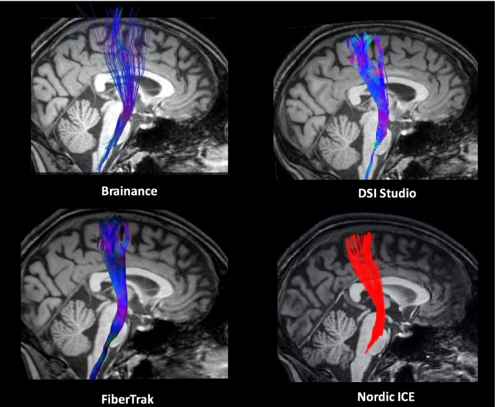
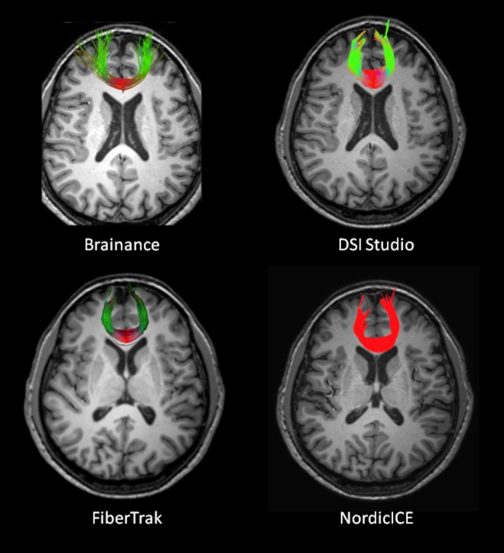
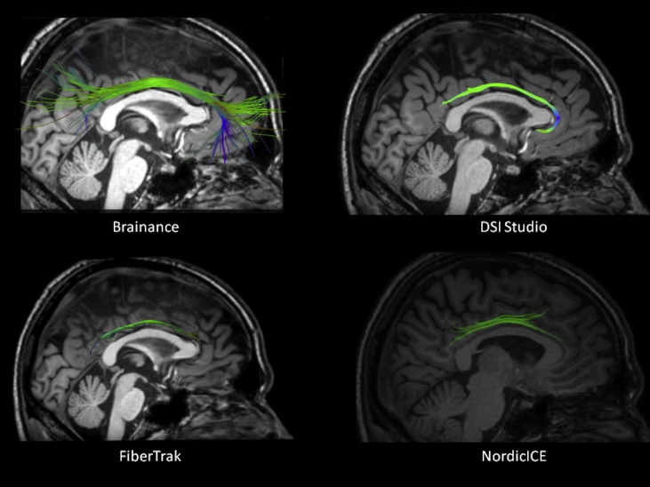
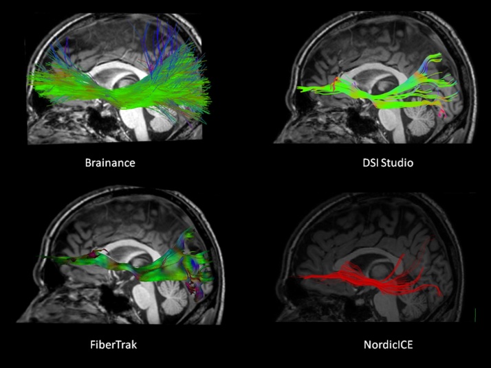
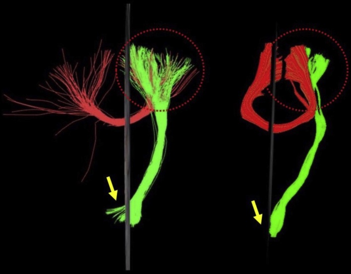
Materials and methods: Ten healthy participants underwent 30-directional diffusion tensor imaging on a 3T-Philips Achieva TX MR-scanner. All data were analyzed by two independent sites of experienced raters with the aforementioned software and the following WM tracts were reconstructed: corticospinal tract (CST); forceps major (Fmajor); forceps minor (Fminor); cingulum bundle (CB); superior longitudinal fasciculus (SLF); inferior fronto-occipital fasciculus (IFOF). Visual inspection of the resulted tracts and statistical analysis (inter-rater and betweensoftware agreement; paired t-test) on fractional anisotropy (FA), axial and radial diffusivity (Daxial, Dradial) were applied for qualitative and quantitative evaluation of DTI software results.
Results: Qualitative evaluation of the extracted tracts confirmed anatomical landmarks at least for the core part of each tract, even though differences in the number of fibers extracted and the whole tract were evident, especially for the CST, Fmajor, Fminor and SLF. Descriptive values did not deviate from the expected range of values for healthy adult population. Substantial inter-rater agreement (intraclass correlation coefficient [ICC], Bland-Altman analysis) was found for all tracts (ICC; FA: 0.839-0.989, Daxial: 0.704-0.991, Dradial: 0.972-0.993). Low agreement for FA, Daxial and Dradial (ICC; Bland-Altman analysis) and significant paired t-test differences (p < 0.05) were detected regarding between-software agreement.
Conclusions: Qualitative comparison of four different DTI software in addition to substantial inter-rater but poor between-software agreement highlight the differences on existing fiber tracking methodologies and several particularities of each WM tract, further supporting the need for further study in both clinical and research settings.
Keywords: Deterministic tractography; Diffusion tensor imaging; Fiber tracking; Magnetic resonance imaging.
Figures
References
-
- Le Bihan D., Breton E., Lallemand D., Grenier P., Cabanis E., Laval-Jeantet M. MR imaging of intravoxel incoherent motions: application to diffusion and perfusion in neurologic disorders. Radiology. 1986;61(2):401–407. - PubMed
-
- Basser P.J., Mattiello J., LeBihan D. Estimation of the effective self-diffusion tensor from the NMR spin echo. J. Magn. Reson. B. 1994;103:247–254. - PubMed
-
- Pierpaoli C., Basser P.J. Toward a quantitative assessment of diffusion anisotropy. Magn. Reson. Med. 1996;36:893–906. - PubMed
-
- Basser P.J., Pajevic S., Pierpaoli C., Duda J., Aldroubi A. In vivo fiber tractography using DT-MRI data. Magn. Reson. Med. 2000;44(4):625–632. - PubMed
Publication types
Grants and funding
LinkOut - more resources
Full Text Sources
Other Literature Sources
