. 2016 Jul 21;1(11):e87632.
doi: 10.1172/jci.insight.87632.

Soluble ADAM33 initiates airway remodeling to promote susceptibility for allergic asthma in early life

Affiliations

Soluble ADAM33 initiates airway remodeling to promote susceptibility for allergic asthma in early life

Elizabeth R Davies et al. JCI Insight. .

Abstract

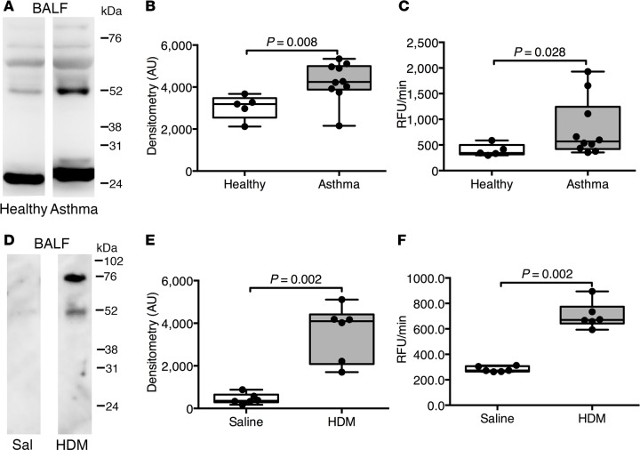
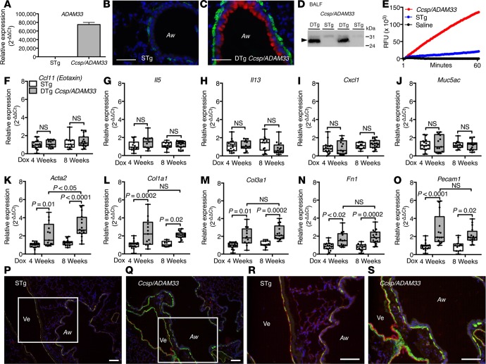
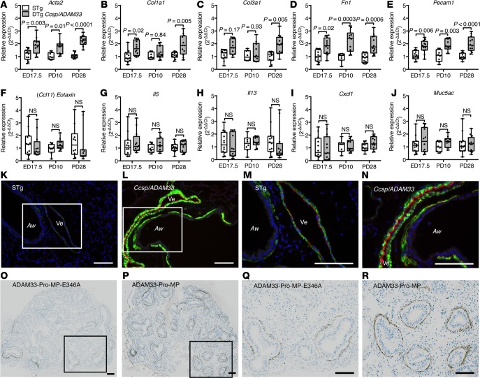
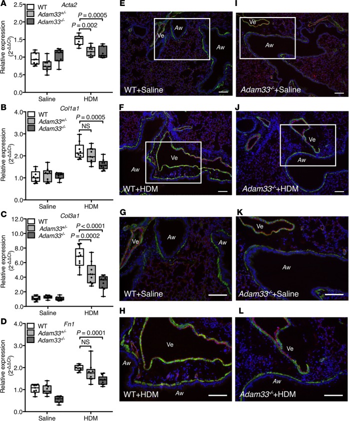
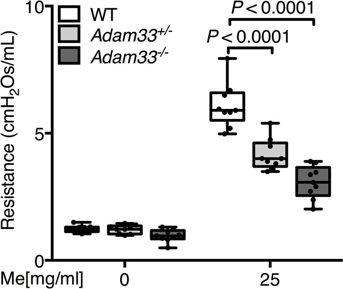
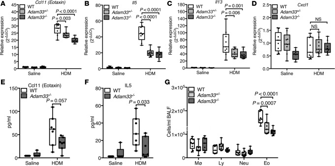
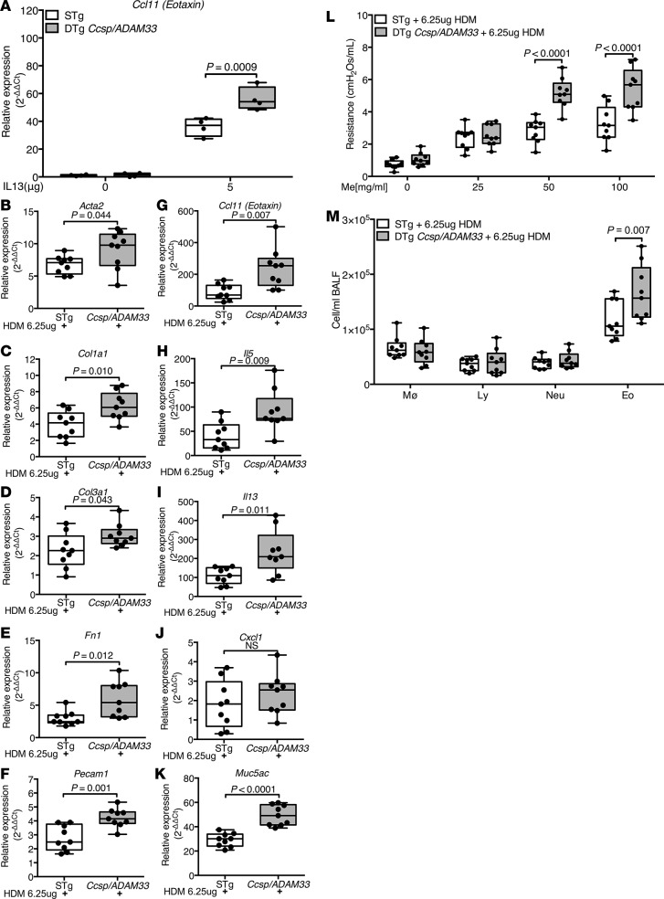
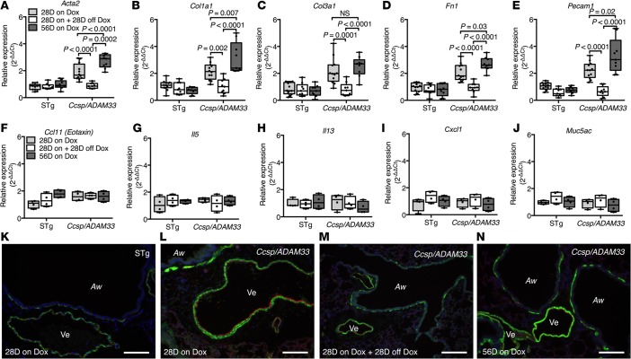
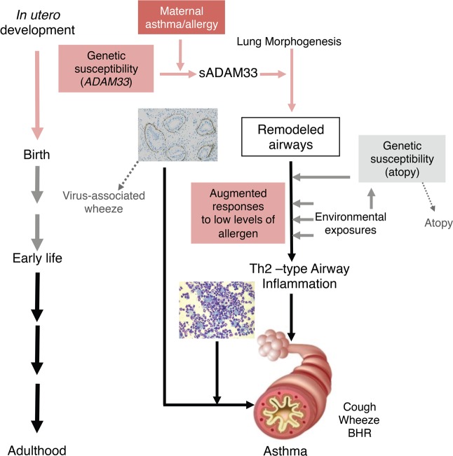
Asthma is a chronic inflammatory airways disease that usually begins in early life and involves gene-environment interactions. Although most asthma exhibits allergic inflammation, many allergic individuals do not have asthma. Here, we report how the asthma gene a disintegrin and metalloprotease 33 (ADAM33) acts as local tissue susceptibility gene that promotes allergic asthma. We show that enzymatically active soluble ADAM33 (sADAM33) is increased in asthmatic airways and plays a role in airway remodeling, independent of inflammation. Furthermore, remodeling and inflammation are both suppressed in Adam33-null mice after allergen challenge. When induced in utero or added ex vivo, sADAM33 causes structural remodeling of the airways, which enhances postnatal airway eosinophilia and bronchial hyperresponsiveness following subthreshold challenge with an aeroallergen. This substantial gene-environment interaction helps to explain the end-organ expression of allergic asthma in genetically susceptible individuals. Finally, we show that sADAM33-induced airway remodeling is reversible, highlighting the therapeutic potential of targeting ADAM33 in asthma.


Figures

Figure 1
Figure 1. Increased soluble ADAM33 (sADAM33) enzymatic activity in bronchoalveolar lavage fluid (BALF) in human asthma and allergic mice.
(A) Western blotting of BALF proteins from healthy (n = 5) and asthmatic (n = 10) subjects using an antibody recognizing the metalloprotease domain of human ADAM33; representative blots are shown. (B) Combined ADAM33-immunoreactive bands (at approximately 25 kDa and between approximately 52 and 76 kDa) were analyzed by densitometry in arbitrary units (AU) (Mann Whitney test). (C) Fluorescence resonance energy transfer (FRET) peptide cleavage assay for ADAM33-specific enzymatic activity in BALF from healthy (n = 5) and asthmatic donors (n = 10), expressed as relative fluorescence units per minute (RFU/min) (Mann Whitney test). (D) Immunoblotting of BALF protein from WT mice challenged with house dust mite (HDM) extract or saline (Sal) (n = 6 per group) with an antibody against mouse ADAM33 extracellular domain. (E) Semiquantitative analysis (bands at approximately 52 and 76 kDa) by densitometry (Mann Whitney test). (F) FRET peptide cleavage assay using murine BALF from HDM-challenged mice or saline controls (n = 6 per group) (Mann Whitney test). Box plots show medians and 25th to 75th percentiles, and whiskers represent minimum and maximum values; all data points are shown. Results are from 3 independent experiments (DF). Full unedited Western blots are available in the Supplemental Material.
Figure 2
Figure 2. Transgenic expression of human soluble ADAM33 (sADAM33) causes airway remodeling.
(A) Human ADAM33 mRNA in single-transgenic (STg) littermate control and double-transgenic (DTg) Ccsp/ADAM33 mouse lungs. (B and C) Representative immunofluorescence staining images for human ADAM33 (red), ACTA2/αSMA (green), and nuclei (blue) in lungs from (B) STg littermate control and (C) DTg Ccsp/ADAM33 mice. Scale bar: 50 μm. (D) Western blotting for human ADAM33 in BALF from DTg Ccsp/ADAM33 or STg control mice. (E) Fluorescence resonance energy transfer (FRET) peptide cleavage assay for ADAM33 enzymatic activity in bronchoalveolar lavage fluid (BALF) from DTg Ccsp/ADAM33 (red) mice, STg (blue) mice, or saline controls (black). Representative traces are shown. (F–O) Relative mRNA expression in whole-lung lobe lysates from adult DTg Ccsp/ADAM33 mice (gray bars) after induction of human ADAM33 for 4 (n = 13) or 8 weeks (n = 16) versus STg littermate controls (white bars) (n = 16 or n = 12, respectively): (F) Ccl11, (G) Il5, (H) Il13, (I) Cxcl1, (J) Muc5ac, (K) Acta2, (L) Col1a1, (M) Col3a1, (N) Fn1, and (O) Pecam1 (2-way ANOVA, Tukey’s multiple comparison test). Box plots show medians and 25th to 75th percentiles, and whiskers represent minimum and maximum values; all data points are shown. Results are from 3 independent experiments (F–O). Representative immunofluorescence staining for ACTA2/αSMA (green), PECAM1 (red), and nuclei (blue) in lungs from (P and R) STg littermate control or (Q and S) DTg Ccsp/ADAM33 mice after 8 weeks of transgene expression. White rectangles in P and Q are shown at higher magnification in R and S. Aw, airway; Ve, vessel. Scale bar: 100 μm. Results are representative of 3 independent experiments (P–S). Full unedited Western blots are available in the Supplemental Material.
Figure 3
Figure 3. Human soluble ADAM33 (sADAM33) causes airway “remodeling” in developing lungs.
(A–J) Reverse-transcription quantitative PCR (RT-qPCR) for remodeling and inflammatory gene expression in whole-lung lobe lysates from double-transgenic (DTg) Ccsp/ADAM33 or single-transgenic (STg) littermate control mice at embryonic day 17.5 (ED17.5) (n = 12/12) and 10 days postpartum (PD10) (n = 8/10) and 28 days postpartum (PD28) (n = 12/12) in which transgene expression was induced by feeding mice doxycycline during pregnancy and up to 4 weeks after birth: (A) Acta2, (B) Col1a1, (C) Col3a1, (D) Fn1 (E) Pecam1 (F) Ccl11, (G) Il5, (H) Il13, (I) Cxcl1, and (J) Muc5ac (2-way ANOVA, Tukey’s multiple comparison test). Box plots show medians and 25th to 75th percentiles, and whiskers represent minimum and maximum values; all data points are shown. Representative immunofluorescence staining for ACTA2/αSMA (green), PECAM1 (red), and nuclei (blue) in lungs from (K and M) STg littermate control or (L and N) DTg Ccsp/Adam33 mice after ADAM33 transgene expression for 4 weeks postpartum. White rectangles in K and L are shown at higher magnification in M and N. Aw, airway; Ve, vessel. Representative immunohistochemistry staining for ACTA2/αSMA (brown) in sections of human embryonic lung explants cultures from 8 to 10 weeks after conception (n = 3) in the presence of (O and Q) recombinant inactive mutant (E346A) ADAM33-Pro-metalloprotease (ADAM33-Pro-MP) and (P and R) enzymatically active ADAM33-Pro-MP. Black rectangles in O and P are shown at higher magnification in Q and R. Scale bar: 100 μm. Results are representative of 3 independent experiments.
Figure 4
Figure 4. Suppression of house dust mite (HDM) extract-induced airway remodeling in Adam33–/– mice.
(A–D) Reverse-transcription quantitative PCR (RT-qPCR) using whole-lung lysates from WT, heterozygote (Adam33+/-), and Adam33–/– mice challenged with saline or HDM extract: (A) Acta2, (B) Col1a1, (C) Col3a1, and (D) Fn1 (n = 9 per group; 2-way ANOVA Tukey’s multiple comparison test). (E–L) Representative immunofluorescence staining for ACTA2/αSMA (green), PECAM1 (red), and nuclei (blue) in tissue sections from mouse lungs after in vivo challenge with saline or HDM extract: (E and G) WT+saline, (F and H) WT+HDM, (I and K) Adam33–/–+saline, and (J and L) Adam33–/–+HDM. White rectangles in E, F, I, and J are shown in G, H, K, and L at higher magnification. Aw, airway; Ve, vessel. Scale bar: 100 μm. Results are representative of 3 independent experiments (E–L).
Figure 5
Figure 5. Suppression of house dust mite (HDM) extract-induced airway hyperresponsiveness in Adam33–/– mice.
Airway resistance in response to methacholine (Me) in WT (white), Adam33+/- (light gray), and Adam33–/– (dark gray) mice following HDM exposure (n = 9 per group; 2-way ANOVA, Tukey’s multiple comparison test). Results are representative of 3 independent experiments.
Figure 6
Figure 6. Suppression of house dust mite (HDM) extract-induced airway inflammation in Adam33–/– mice.
(A–D) Reverse-transcription quantitative PCR (RT-qPCR) using whole-lung lysates from WT (white), heterozygote (Adam33+/-) (light gray), and Adam33–/– (dark gray) mice challenged with saline or HDM extract: (A) Ccl11, (B) Il5, (C) Il13, and (D) Cxcl1; (n = 9 per group; 2-way ANOVA Tukey’s multiple comparison test). (E and F) Multiplex assay for CCL11 and IL-5 protein levels in bronchoalveolar lavage fluid (BALF) (n = 5 or 7 per group; 1-way ANOVA, Tukey’s multi comparison test). (G) Differential inflammatory cell counts for macrophages (MØ), lymphocytes (Ly), neutrophils (Neu), and eosinophils (Eo) in BALF from WT, Adam33+/-, and Adam33–/– mice challenged with HDM (n = 9 per group; 2-way ANOVA Tukey’s multiple comparison test). Box plots show medians and 25th to 75th percentiles, and whiskers minimum to maximum; all data points are shown. Results are from 3 independent experiments.
Figure 7
Figure 7. Soluble ADAM33 (sADAM33) augments airway responses to allergens.
(A) Reverse-transcription quantitative PCR (RT-qPCR) for Ccl11 mRNA expression in lung lobe lysates 24 hours after intratracheal installation of 5.0 μg murine IL-13 in double-transgenic (DTg) Ccsp/ADAM33 (gray bars) and single-transgenic (STg) littermate control mice (white bars) after transgene induction for 4 weeks (n = 4/group; 2-way ANOVA, Tukey’s multiple comparison test). Results are from 1 experiment. (B–M) After transgene induction for 6 weeks, DTg Ccsp/ADAM33 or STg control mice were sensitized and challenged with house dust mite (HDM) extract (6.5 μg) for analysis of gene expression, bronchial hyperresponsiveness (BHR), and inflammation. (B–K) RT-qPCR for relative mRNA expression (compared with saline-challenged mice) in lung lobe lysates from DTg Ccsp/ADAM33 or STg control mice: (B) Acta2, (C) Col1a1, (D) Col3a1, (E) Fn1, (F) Pecam1, (G) Ccl11, (H) Il5, (I) Il13, (J) Cxcl1, and (K) Muc5ac (all n = 9 per group; unpaired Student’s t test or Mann Whitney test). (L) Airway resistance in response to increasing concentrations of methacholine (Me) and (M) differential inflammatory cell counts for macrophages (MØ), lymphocytes (Ly), neutrophils (Neu), and eosinophils (Eo) in bronchoalveolar lavage fluid (BALF) after HDM or saline challenge (n = 9 per group; 2-way ANOVA, Tukey’s multiple comparison test). Box plots show medians and 25th to 75th percentiles, and whiskers represent minimum and maximum values; all data points are shown. Results are from 3 independent experiments.
Figure 8
Figure 8. Airway remodeling induced by soluble ADAM33 (sADAM33) is reversible.
(A–J) Reverse-transcription quantitative PCR (RT-qPCR) for remodeling (n = 9/group) and inflammatory (n = 4/group) gene expression in whole-lung lobe lysates from double-transgenic (DTg) Ccsp/ADAM33 or single-transgenic (STg) littermate control mice in which transgene expression was induced by doxycycline (Dox) feeding during pregnancy and for up to 28 days (28D on Dox) or 56 days (56D on Dox) after birth or Dox feeding for 28 days after birth followed by a cessation of Dox for 28 days (28D on + 28D off Dox): (A) Acta2, (B) Col1a1, (C) Col3a1, (D) Fn1 (E) Pecam1, (F) Ccl11, (G) Il5, (H) Il13, (I) Cxcl1, and (J) Muc5ac (2-way ANOVA, Tukey’s multiple comparison test). Box plots show medians and 25th to 75th percentiles, and whiskers represent minimum and maximum values; all data points are shown. Results are from 3 independent experiments (A–E) and from 1 experiment (F–J). (K–N) Representative immunofluorescence staining for ACTA2/αSMA (green), PECAM1 (red), and nuclei (blue) in lungs from (K) STg littermate control or (L) DTg Ccsp/Adam33 mice in which transgene expression was induced in utero and by Dox feeding postpartum for 28 days and (N) after 56 days. (M) DTg Ccsp/Adam33 mice after ADAM33 transgene expression was induced in utero and by 28 days of Dox feeding postpartum and then 28 days off Dox. Aw, airway; Ve, vessel. Scale bar: 100 μm. Results are representative of 2 independent experiments (K–N).
Figure 9
Figure 9. Schematic representation of the contribution of soluble ADAM33 (sADAM33) as a local tissue susceptibility gene in asthma pathobiology.

References

    1. Cookson WO, Moffatt MF. Genetics of asthma and allergic disease. Hum Mol Genet. 2000;9(16):2359–2364. doi: 10.1093/hmg/9.16.2359. - DOI - PubMed
    1. Kim HY, DeKruyff RH, Umetsu DT. The many paths to asthma: phenotype shaped by innate and adaptive immunity. Nat Immunol. 2010;11(7):577–584. doi: 10.1038/ni.1892. - DOI - PMC - PubMed
    1. Lambrecht BN, Hammad H. The immunology of asthma. Nat Immunol. 2015;16(1):45–56. - PubMed
    1. Fahy JV. Type 2 inflammation in asthma--present in most, absent in many. Nat Rev Immunol. 2015;15(1):57–65. - PMC - PubMed
    1. Martinez FD, Vercelli D. Asthma. Lancet. 2013;382(9901):1360–1372. doi: 10.1016/S0140-6736(13)61536-6. - DOI - PMC - PubMed