. 2016 Sep;590(18):3168-78.
doi: 10.1002/1873-3468.12341. Epub 2016 Sep 12.

N-glycosylation in the thermoacidophilic archaeon Sulfolobus acidocaldarius involves a short dolichol pyrophosphate carrier

Affiliations

N-glycosylation in the thermoacidophilic archaeon Sulfolobus acidocaldarius involves a short dolichol pyrophosphate carrier

Ziqiang Guan et al. FEBS Lett. 2016 Sep.

Abstract

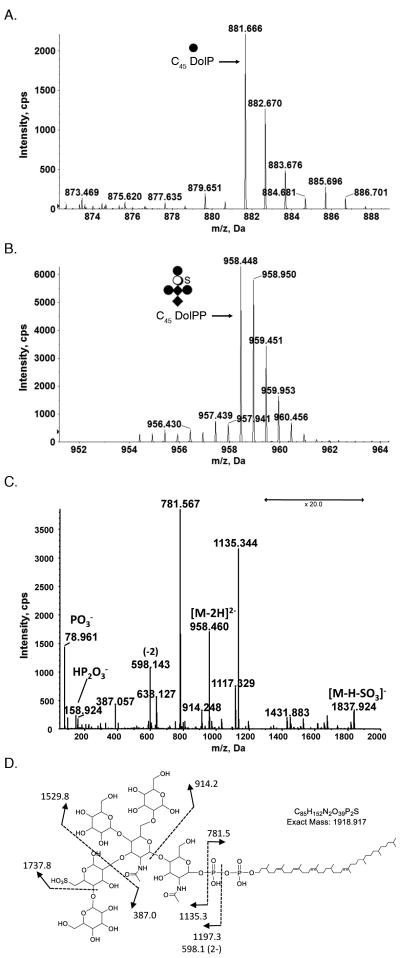
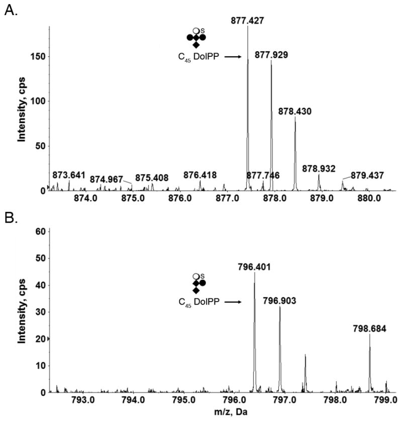
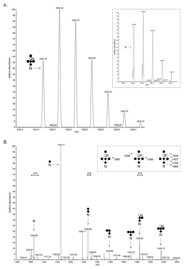
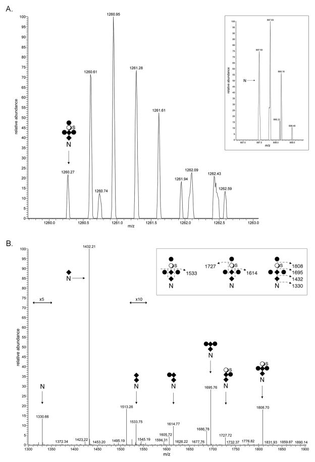
N-glycosylation is a post-translational modification that occurs across evolution. In the thermoacidophilic archaea Sulfolobus acidocaldarius, glycoproteins are modified by an N-linked tribranched hexasaccharide reminiscent of the N-glycans assembled in Eukarya. Previously, hexose-bearing dolichol phosphate was detected in a S. acidocaldarius Bligh-Dyer lipid extract. Here, we used a specialized protocol for extracting lipid-linked oligosaccharides to detect a dolichol pyrophosphate bearing the intact hexasaccharide, as well as its biosynthetic intermediates. Furthermore, evidence for N-glycosylation of two S. acidocaldarius proteins by the same hexasaccharide and its derivatives was collected. These findings thus provide novel insight into archaeal N-glycosylation.

Keywords: N-glycosylation; Sulfolobus acidocaldarius; archaea; dolichol; thermoacidophile.


Figures

Fig 1
Fig 1
Glycan-charged phosphorylated dolichol species in S. acidocaldarius. LC-ESI MS analysis of a S. acidocaldarius lipid extract revealed the presence of (A) C45 DolP modified by a hexose and (B) C45 DolPP modified by a hexasaccharide comprising N-acetylhexosamine2-hexose3-sulfated quinovose (QuiS). C. MS/MS spectrum of [M-2H]2− at m/z 958.4. D. Fragmentation scheme for the observed product ions in C. The depicted DolPP-linked glycan structure is based on the previously reported protein N-linked glycan [24,25]. The arrows indicating x20 reflect magnification of the ion peaks in the corresponding region of the m/z values on the graph. Where the position of a double bond is only speculated, a dotted line is drawn. Diamonds represent N-acetylhexosamines, full circles represent hexoses, open circles represent quinovose and S represents sulfation.
Fig 2
Fig 2
Glycan-charged S. acidocaldarius DolPP species. (A) C45 DolP modified by a pentasaccharide comprising N-acetylhexosamine2-hexose2-sulfated quinovose. (B) C45 DolP modified by a tetrasaccharide comprising N-acetylhexosamine2-hexose-sulfated quinovose. Diamonds represent N-acetylhexosamines, full circles represent hexoses, open circles represent quinovose and S represents sulfation.
Fig 3
Fig 3
N-glycosylation of S. acidocaldarius SlaB. A. LC-ESI MS analysis of a tryptic fragment of SlaB that includes Asn-278 revealed the peptide (inset) and its modification by a hexasaccharide comprising N-acetylhexosamine2-hexose3-sulfated quinovose. [M+2H]2+ ion peaks are presented. B. MS/MS profile of the [M+2H]2+ base peak of the peptide observed at m/z 1621.72. The inset schematically represents the fragmentation scheme. The N represents Asn-278, diamonds represent N-acetylhexosamines, full circles represent hexoses, open circles represent quinovose and S represents sulfation. The arrows indicating x10 reflect magnification of the ion peaks in the corresponding region of the m/z values on the graph.
Fig 4
Fig 4
N-glycosylation of S. acidocaldarius FlaB. A. LC-ESI MS analysis of a tryptic fragment of FlaB that includes Asn-195 revealed the peptide (inset) and its modification by a hexasaccharide comprising N-acetylhexosamine2-hexose3-sulfated quinovose. [M+3H]3+ ion peaks are presented. B. MS/MS profile of the 13C1-containing isotopic [M+2H]2+ peak at m/z 1890.291. The inset schematically represents the fragmentation scheme. The N represents Asn-195, diamonds represent N-acetylhexosamines, full circles represent hexoses, open circles represent quinovose and S represents sulfation. The arrows indicating x5 and x10 reflect magnification of the ion peaks in the corresponding region of the m/z values on the graph.

References

    1. Swiezewska E, Danikiewicz W. Polyisoprenoids: structure, biosynthesis and function. Prog Lipid Res. 2005;44:235–258. - PubMed
    1. Jones MB, Rosenberg JN, Betenbaugh MJ, Krag SS. Structure and synthesis of polyisoprenoids used in N-glycosylation across the three domains of life. Biochim Biophys Acta. 2009;1790:485–494. - PMC - PubMed
    1. Guan Z, Eichler J. Liquid chromatography/tandem mass spectrometry of dolichols and polyprenols, lipid sugar carriers across evolution. Biochim Biophys Acta. 2011;1811:800–806. - PMC - PubMed
    1. Hartley MD, Imperiali B. At the membrane frontier: a prospectus on the remarkable evolutionary conservation of polyprenols and polyprenyl-phosphates. Arch Biochem Biophys. 2012;517:83–97. - PMC - PubMed
    1. Eichler J. Extreme sweetness: protein glycosylation in Archaea. Nat Rev Microbiol. 2013;11:151–156. - PubMed

Publication types

MeSH terms

LinkOut - more resources