. 2016 Aug;8(8):1703-17.
doi: 10.18632/aging.101007.

Paracrine effects of human adipose-derived mesenchymal stem cells in inflammatory stress-induced senescence features of osteoarthritic chondrocytes

Affiliations

Paracrine effects of human adipose-derived mesenchymal stem cells in inflammatory stress-induced senescence features of osteoarthritic chondrocytes

Julia Platas et al. Aging (Albany NY). 2016 Aug.

Abstract

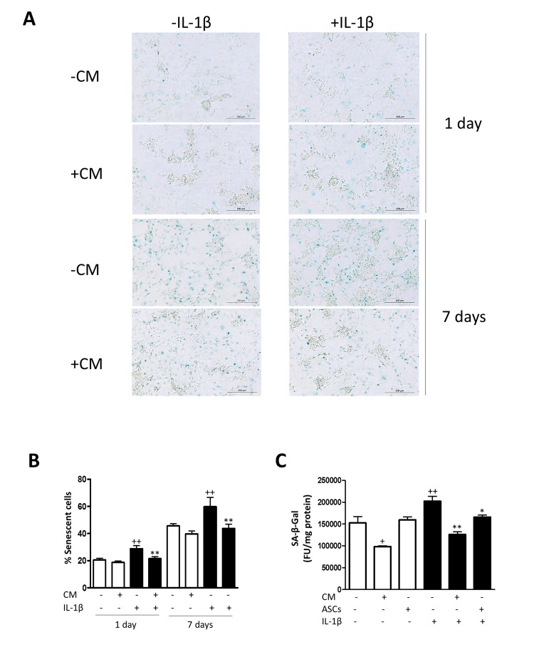
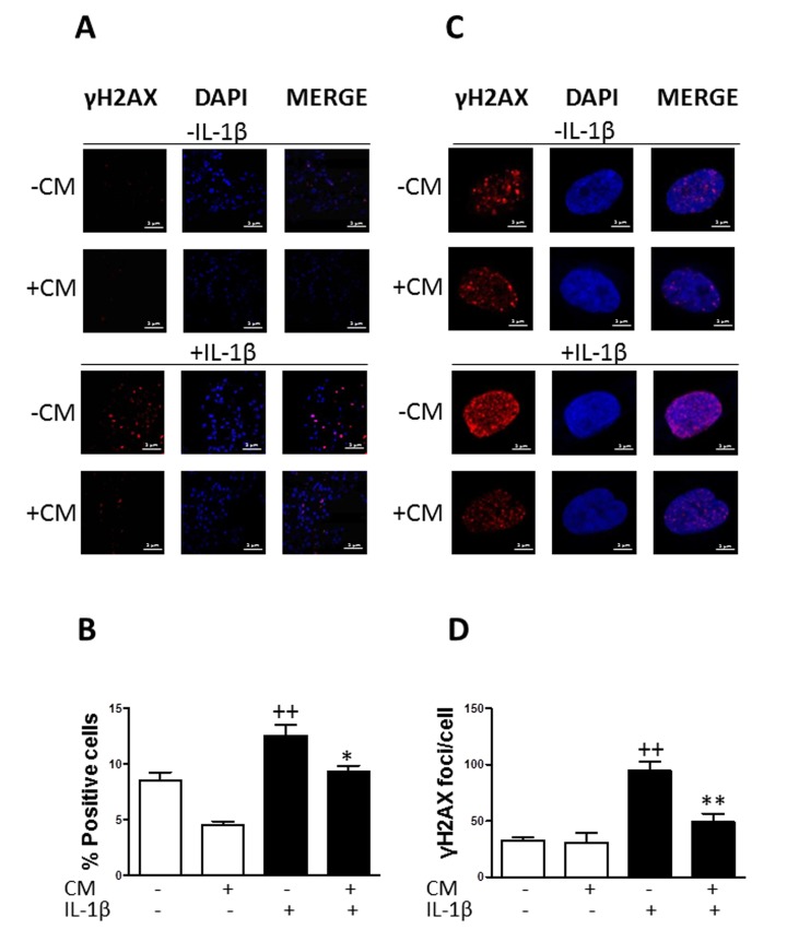
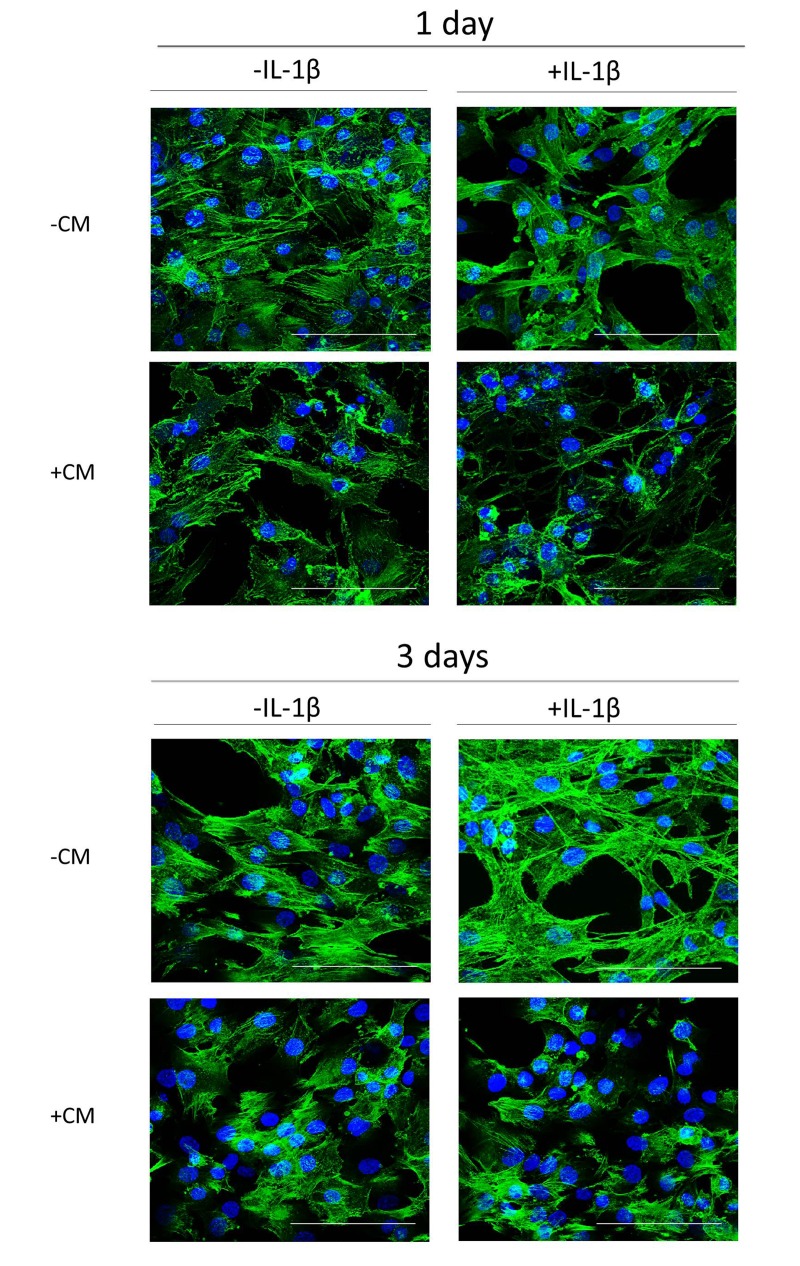
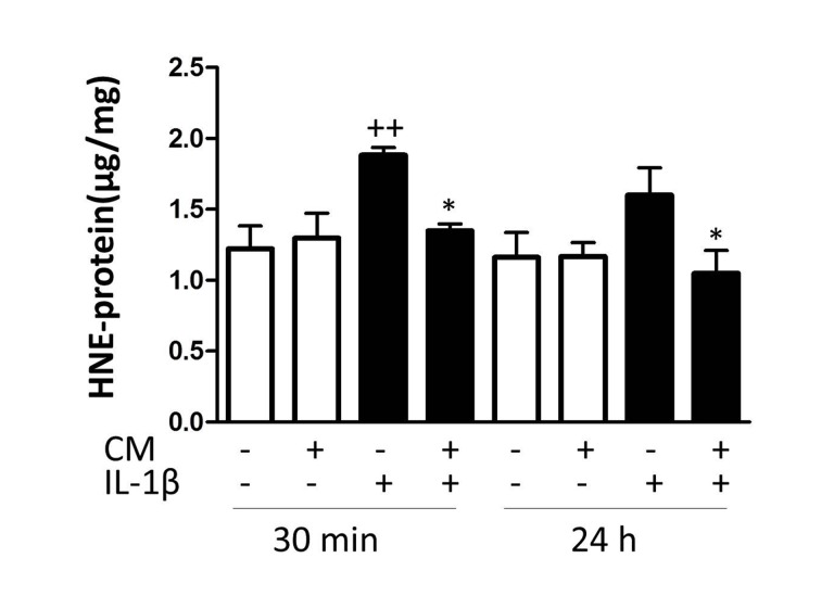
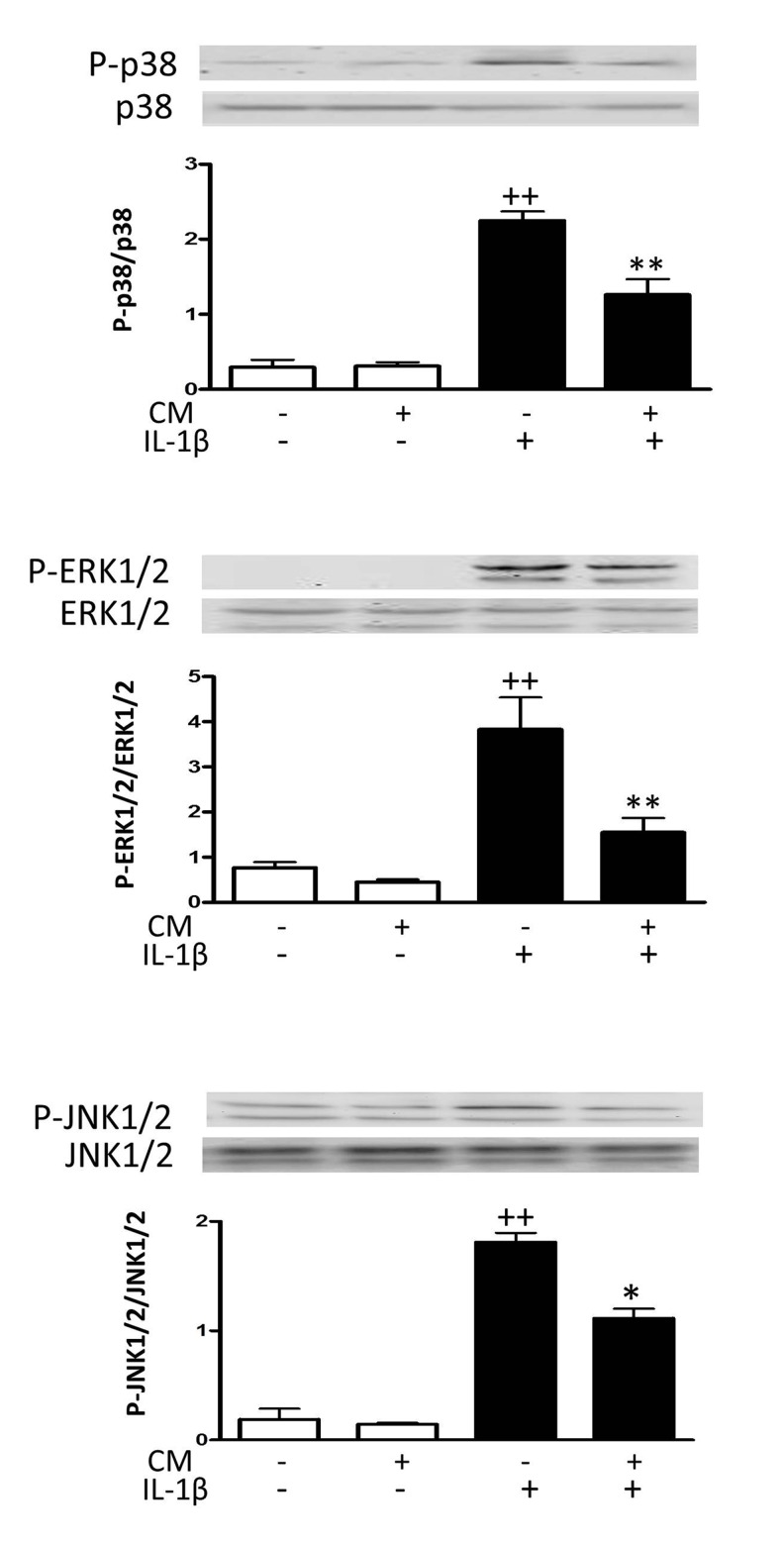
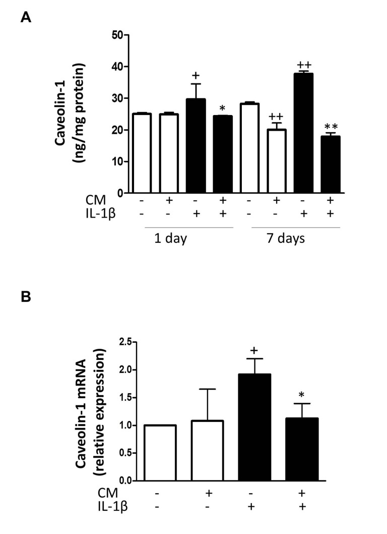
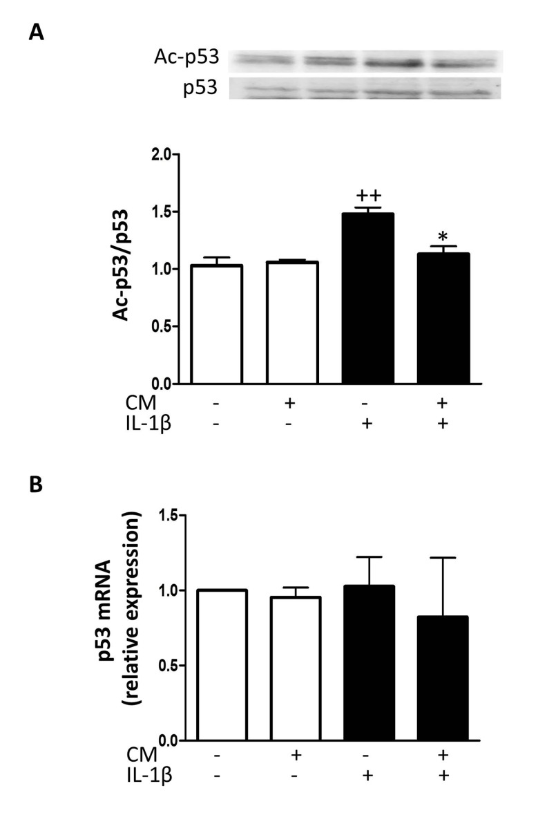
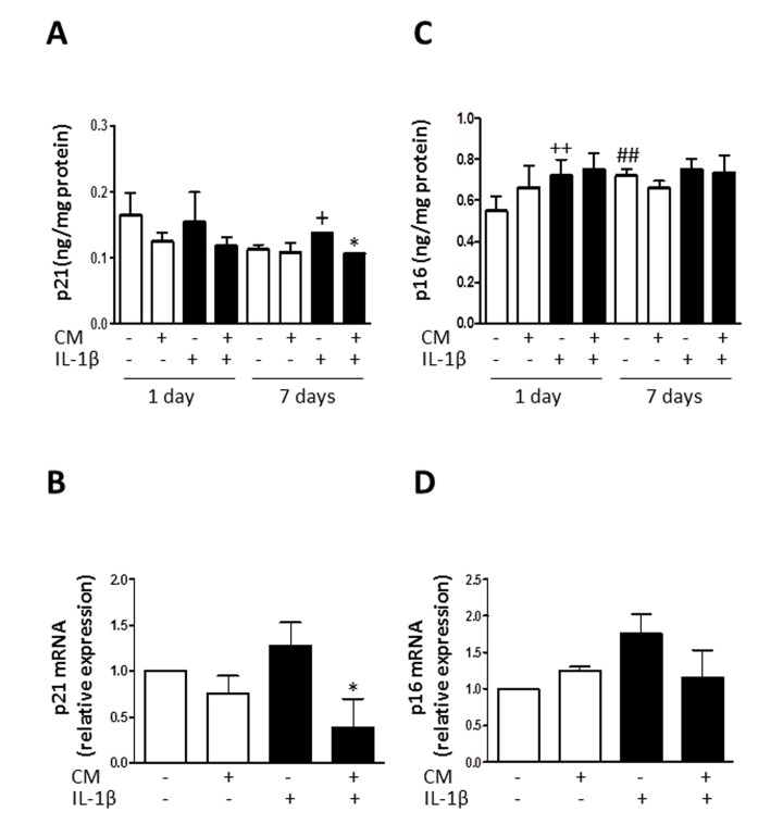
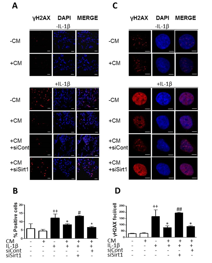
Aging and exposure to stress would determine the chondrocyte phenotype in osteoarthritis (OA). In particular, chronic inflammation may contribute to stress-induced senescence of chondrocytes and cartilage degeneration during OA progression. Recent studies have shown that adipose-derived mesenchymal stem cells exert paracrine effects protecting against degenerative changes in chondrocytes. We have investigated whether the conditioned medium (CM) from adipose-derived mesenchymal stem cells may regulate senescence features induced by inflammatory stress in OA chondrocytes. Our results indicate that CM down-regulated senescence markers induced by interleukin-1β including senescence-associated β-galactosidase activity, accumulation of γH2AX foci and morphological changes with enhanced formation of actin stress fibers. Treatment of chondrocytes with CM also decreased the production of oxidative stress, the activation of mitogen-activated protein kinases, and the expression of caveolin-1 and p21. The effects of CM were related to the reduction in p53 acetylation which would be dependent on the enhancement of Sirtuin 1 expression. Therefore, CM may exert protective effects in degenerative joint conditions by countering the premature senescence of OA chondrocytes induced by inflammatory stress.

Keywords: adipose-derived mesenchymal stem cells conditioned medium; chondrocytes; inflammation; senescence.


Conflict of interest statement

CONFLICT OF INTERESTS The authors declare no conflict of interests.

Figures

Figure 1
Figure 1. Induction of SA-β-Gal by IL-1β in OA chondrocytes
(A) Representative light microscopy images of cells stained with the senescent marker SA-β-Gal in monolayer cultures of OA chondrocytes. Bars: 200 μm. (B) Percentage of SA-β-Gal positive cells. OA chondrocytes were incubated with IL-1β and/or CM for 1 day and 7 days. (C) SA-β-Gal was quantified by fluorometry. A co-culture system of AMSC and OA chondrocytes was used. Data are shown as mean±standard deviation of N=4 separate experiments with cells from separate donors. +p<0.05, ++p<0.01 with respect to non-treated cells; *p<0.05, **p<0.01 with respect to IL-1β-treated cells. FU: fluorescence units.
Figure 2
Figure 2. Effect of CM on γ-H2AX foci accumulation induced by IL-1β in OA chondrocytes
(A) Presence of γH2AX (red pixels) positive cells in a representative experiment. DAPI was used to stain the nuclei. Bars: 50μm. (B) Percentage of positive cells. (C) Presence of γH2AX (red pixels) foci in cell nucleus in a representative experiment. DAPI was used to stain the nuclei. Bars: 5μm. (D) Number of γH2AX foci per cell. OA chondrocytes were incubated with IL-1β and/or CM for 3 days. Data are shown as mean±standard deviation of N=4 separate experiments with cells from separate donors. ++p<0.01 with respect to non-treated cells; *p<0.05, **p<0.01, with respect to IL-1β-treated cells.
Figure 3
Figure 3. Actin stress fibers formation induced by IL-1β in OA chondrocytes and effect of CM
Presence of actin stress fibers (green pixels) in a representative experiment of N=4 separate experiments with cells from separate donors. DAPI was used to stain the nuclei. Bars: 50 μm. OA chondrocytes were incubated with IL-1β and/or CM for 1 day and 3 days.
Figure 4
Figure 4. Effect of CM on oxidative stress induced by IL-1β in OA chondrocytes
The intracellular levels of HNE-modified proteins were quantified by ELISA. OA chondrocytes were incubated with IL-1β and/or CM for the indicated times. Data are shown as mean±standard deviation of N=9 separate experiments with cells from separate donors. ++p<0.01 with respect to non-treated cells; *p<0.05, with respect to IL-1β-treated cells.
Figure 5
Figure 5. Effect of CM on MAPK
OA chondrocytes were incubated with IL-1β and/or CM for 15 min and immunoblotting was performed for phosphorylated and total p38, ERK1/2 and JNK1/2. Data are shown as mean±standard deviation of N=3 separate experiments with cells from separate donors. ++p<0.01 with respect to non-treated cells; *p<0.05, **p<0.01 with respect to IL-1β-treated cells.
Figure 6
Figure 6. Effect of CM on caveolin-1
(A) Protein levels of caveolin-1 were assessed by ELISA. OA chondrocytes were incubated with IL-1β and/or CM for the indicated times. (B) Caveolin-1 mRNA expression was determined by quantitative real-time PCR. OA chondrocytes were incubated with IL-1β and/or CM for 24 hours. Data are shown as mean±standard deviation of N=4 separate experiments with cells from separate donors. +p<0.05, ++p<0.01 with respect to non-treated cells; *p<0.05, **p<0.01 with respect to IL-1β-treated cells.
Figure 7
Figure 7. Effect of CM on p53
(A) Immunoblotting was performed for acetylated and total p53. (B) p53 mRNA expression was determined by quantitative real-time PCR. OA chondrocytes were incubated with IL-1β and/or CM for 24 hours. Data are shown as mean±standard deviation of N=4 separate experiments with cells from separate donors. ++p<0.01 with respect to non-treated cells; *p<0.05 with respect to IL-1β-treated cells.
Figure 8
Figure 8. Effect of CM on p21 and p16
(A) Protein levels of p21 were assessed by ELISA. OA chondrocytes were incubated with IL-1β and/or CM for the indicated times. (B) p21 mRNA expression was determined by quantitative real-time PCR. OA chondrocytes were incubated with IL-1β and/or CM for 24 hours. (C) Protein levels of p16 were assessed by ELISA. OA chondrocytes were incubated with IL-1β and/or CM for the indicated times. (D) p16 mRNA expression was determined by quantitative real-time PCR. OA chondrocytes were incubated with IL-1β and/or CM for 24 hours. Data are shown as mean±standard deviation of N=8 separate experiments with cells from separate donors. +p<0.05, ++p<0.01 with respect to non-treated cells; *p<0.05 with respect to IL-1β-treated cells; ##p<0.01 with respect to non-treated cells at day 1.
Figure 9
Figure 9. Effect of CM on Sirt1
(A) Protein levels of Sirt1 were assessed by ELISA. OA chondrocytes were incubated with IL-1β and/or CM for the indicated times. (B) Sirt1 mRNA expression was determined by quantitative real-time PCR. OA chondrocytes were incubated with IL-1β and/or CM for 24 hours. Data are shown as mean±standard deviation of N=8 separate experiments with cells from separate donors. +p<0.05, ++p<0.01 with respect to non-treated cells; *p<0.05, **p<0.01 with respect to IL-1β-treated cells.
Figure 10
Figure 10. Effect of Sirt1 siRNA on the reduction of γH2AX foci accumulation by CM
(A) Representative experiment showing that Sirt1 siRNA (siSirt1) inhibits the effect of CM on the accumulation of γH2AX (red pixels) positive cells induced by IL-1β. siCont: non-specific siRNA. DAPI was used to stain the nuclei. Bars: 50μm. (B) Percentage of positive cells. (C) Representative experiment showing that Sirt1 siRNA (siSirt1) inhibits the effect of CM on the accumulation of γH2AX (red pixels) foci per cell induced by IL-1β. siCont: non-specific siRNA. DAPI was used to stain the nuclei. Bars: 5μm. (D) Number of γH2AX foci per cell. OA chondrocytes were incubated with the indicated treatments for 3 days. Data are shown as mean±standard deviation of N=4 separate experiments with cells from separate donors. ++p<0.01 with respect to non-treated cells; *p<0.05 with respect to IL-1β-treated cells; #p<0.05, ##p<0.01 with respect to IL-1β+CM.

References

    1. Bijlsma JW, Berenbaum F, Lafeber FP. Osteoarthritis: an update with relevance for clinical practice. Lancet. 2011;377:2115–26. - PubMed
    1. Goldring MB, Birkhead J, Sandell LJ, Kimura T, Krane SM. Interleukin 1 suppresses expression of cartilage-specific types II and IX collagens and increases types I and III collagens in human chondrocytes. J Clin Invest. 1988;82:2026–37. - PMC - PubMed
    1. Tetlow LC, Adlam DJ, Woolley DE. Matrix metalloproteinase and proinflammatory cytokine production by chondrocytes of human osteoarthritic cartilage: associations with degenerative changes. Arthritis Rheum. 2001;44:585–94. - PubMed
    1. Martin JA, Buckwalter JA. The role of chondrocyte senescence in the pathogenesis of osteoarthritis and in limiting cartilage repair. J Bone Joint Surg Am. 2003;85-A:106–10. - PubMed
    1. Aigner T, Hemmel M, Neureiter D, Gebhard PM, Zeiler G, Kirchner T, McKenna L. Apoptotic cell death is not a widespread phenomenon in normal aging and osteoarthritis human articular knee cartilage: a study of proliferation, programmed cell death (apoptosis), and viability of chondrocytes in normal and osteoarthritic human knee cartilage. Arthritis Rheum. 2001;44:1304–12. - PubMed

MeSH terms