. 2016 Aug 4:5:e17983.
doi: 10.7554/eLife.17983.

Structures of TorsinA and its disease-mutant complexed with an activator reveal the molecular basis for primary dystonia

Affiliations

Structures of TorsinA and its disease-mutant complexed with an activator reveal the molecular basis for primary dystonia

F Esra Demircioglu et al. Elife. .

Abstract

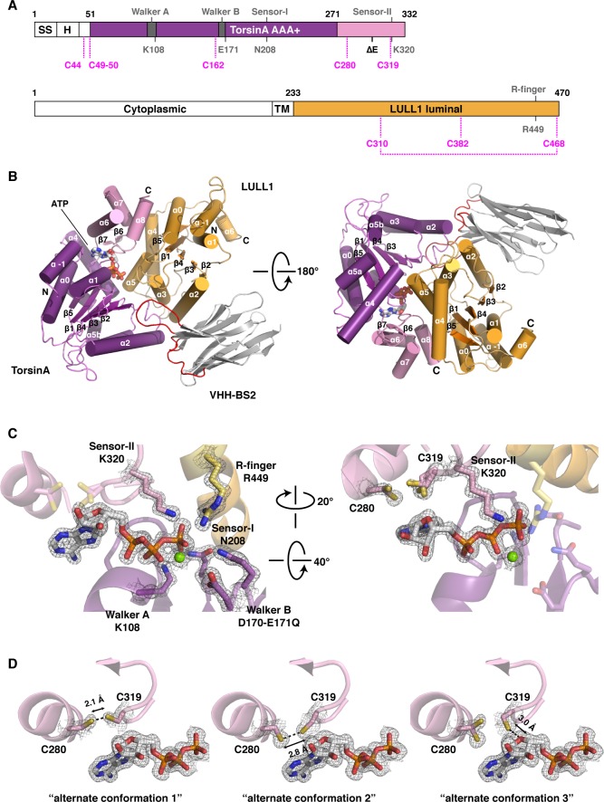
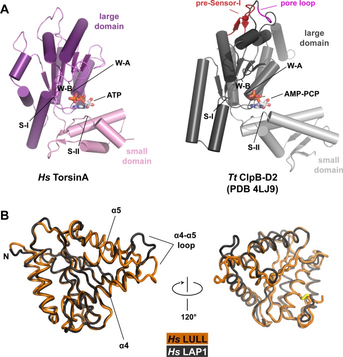
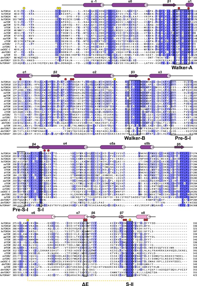
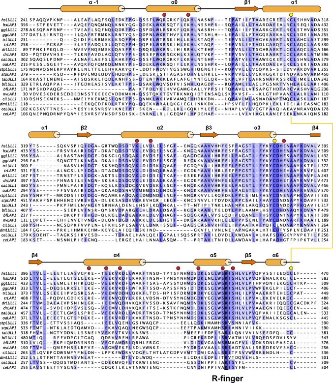
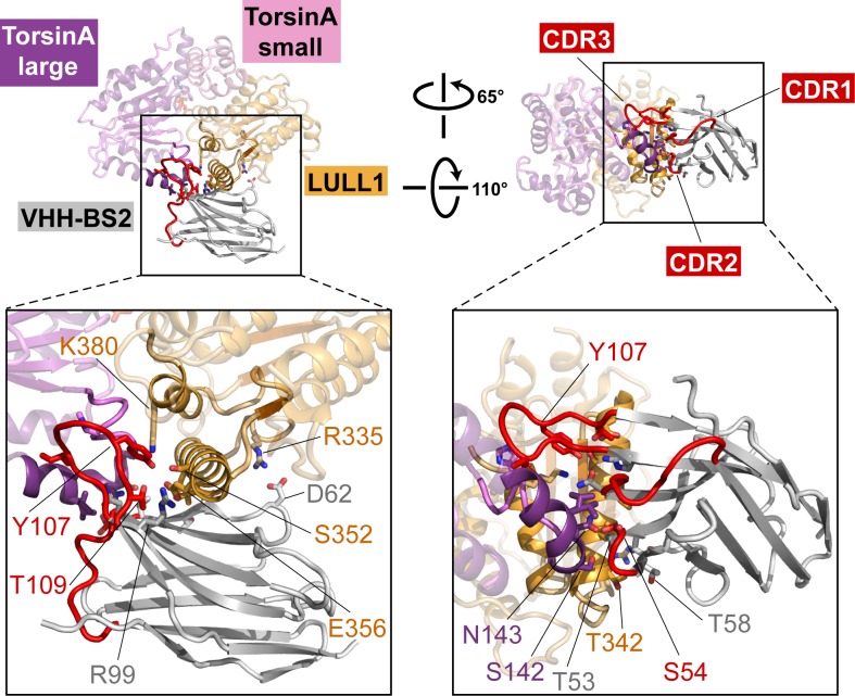
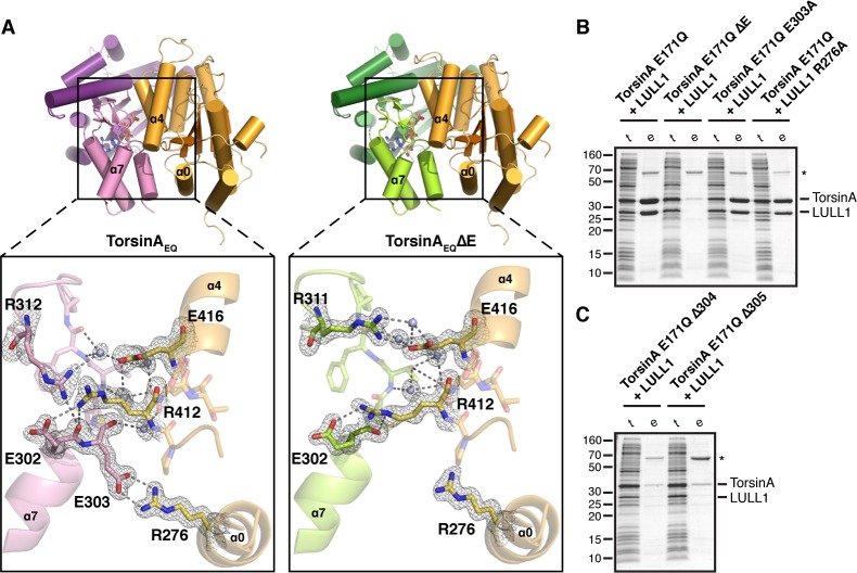
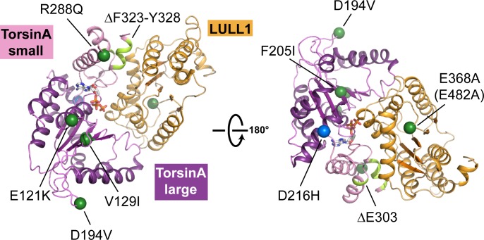
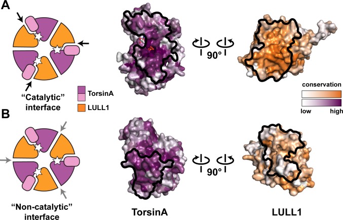
The most common cause of early onset primary dystonia, a neuromuscular disease, is a glutamate deletion (ΔE) at position 302/303 of TorsinA, a AAA+ ATPase that resides in the endoplasmic reticulum. While the function of TorsinA remains elusive, the ΔE mutation is known to diminish binding of two TorsinA ATPase activators: lamina-associated protein 1 (LAP1) and its paralog, luminal domain like LAP1 (LULL1). Using a nanobody as a crystallization chaperone, we obtained a 1.4 Å crystal structure of human TorsinA in complex with LULL1. This nanobody likewise stabilized the weakened TorsinAΔE-LULL1 interaction, which enabled us to solve its structure at 1.4 Å also. A comparison of these structures shows, in atomic detail, the subtle differences in activator interactions that separate the healthy from the diseased state. This information may provide a structural platform for drug development, as a small molecule that rescues TorsinAΔE could serve as a cure for primary dystonia.

Keywords: AAA+ ATPases; biophysics; cell biology; dystonia; human; nuclear envelope; structural biology.


Conflict of interest statement

FED, BAS and TUS: Filed a provisional patent application protecting the use of the crystal structures (U.S.P.T.O. No. 62/330,683). The other authors declare that no competing interests exist.

Figures

Figure 1.
Figure 1.. Architecture of the TorsinA-LULL1 complex.
(A) Schematic diagrams of TorsinA and LULL1. Important residues and sequence motifs are indicated. The colored areas mark the crystallized segments. Large and small domains of TorsinA are colored in purple and pink, respectively. SS, signal sequence; H, hydrophobic region; TM, transmembrane helix. (B) Cartoon representation of the TorsinA-LULL1 complex in two orientations. Color-coding as in (A). A nanobody (VHH-BS2, grey; complementarity determining regions, red) was used as a crystallization chaperone. Numbers refer to secondary structure elements. (C) Close-up of the ATP binding site. Key residues are labeled. 2Fo−Fc electron density contoured at 2σ displayed as grey mesh. (D) Close-up of the proximal cysteines 280 and 319 next to the adenine base of the bound ATP. 2Fo−Fc electron density is contoured at 1σ. The cysteine pair adopts three alternate conformations, but remains reduced in all of them. DOI: http://dx.doi.org/10.7554/eLife.17983.003
Figure 1—figure supplement 1.
Figure 1—figure supplement 1.. Structural comparisons.
(A) Human TorsinA-ATP (left) displayed as a cartoon, compared to the D2 domain of the double-ringed AAA+ ATPase ClpB-AMPPCP (right) from Thermus thermophilus (Zeymer et al., 2014) (PDB code 4LJ9) in the same orientation. Important structure motifs are labeled. (B) Human LULL1 (orange) superposed on human LAP1 (grey, PDB code 4TVS), shown in two orientations. The one region of major structural difference is labeled (left). The disulfide bridge within LAP1/LULL1 is in yellow (right). DOI: http://dx.doi.org/10.7554/eLife.17983.004
Figure 1—figure supplement 2.
Figure 1—figure supplement 2.. Phylogenetic analysis of Torsins.
Maximally diverged torsins are aligned. Secondary structure elements of human TorsinA are displayed above the alignment. Important sequence motifs are boxed. LULL1 contacts, red circles, conserved cysteines, yellow circles. Proximal cysteines 280 and 319 connected with a dashed yellow line. Asterisk denotes putative torsin homologs based on sequence analysis. hs, Homo sapiens; oa, Ornithorhynchus anatinus; gg, Gallus gallus; tr, Takifugu rubripes; dr, Danio rerio; nv, Nematostella vectensis; bf, Branchiostoma floridae; stp, Strongylocentrotus purpuratus; ci, Ciona intestinalis; ce, Caenorhabditis elegans; dm, Drosophila melanogaster; ta, Trichoplax adherens. DOI: http://dx.doi.org/10.7554/eLife.17983.005
Figure 1—figure supplement 3.
Figure 1—figure supplement 3.. Phylogenetic analysis of LAP1/LULL1.
Maximally diverged LAP1 and LULL1 sequences are aligned. If not experimentally confirmed, sequences were assigned as LAP1 or LULL1 based on the presence of an N-terminal, extraluminal domain with basic signature, characteristic of LAP1. Secondary structure elements of human LULL1 are displayed above the alignment. The strictly conserved Arg-finger is boxed. TorsinA contacts, red circles, conserved cysteines, yellow circles. Disulfide bridge depicted as a yellow line. hs, Homo sapiens; oa, Ornithorhynchus anatinus; gg, Gallus gallus; tr, Takifugu rubripes; dr, Danio rerio; nv, Nematostella vectensis; bf, Branchiostoma floridae; stp, Strongylocentrotus purpuratus; ci, Ciona intestinalis; ce, Caenorhabditis elegans; dm, Drosophila melanogaster; ta, Trichoplax adherens. DOI: http://dx.doi.org/10.7554/eLife.17983.006
Figure 1—figure supplement 4.
Figure 1—figure supplement 4.. Nanobody interaction.
The heterotrimeric TorsinA(ATP)-LULL1-VHH-BS2 complex is shown in two orientations. Nanobody and interacting secondary structure elements of TorsinA and LULL1 are shown in full color, non-interacting elements in faded colors. Complementarity determining region (CDR) loops in red. Insets show close-ups with important interacting residues labeled. DOI: http://dx.doi.org/10.7554/eLife.17983.007
Figure 1—figure supplement 5.
Figure 1—figure supplement 5.. Comparison of sequence motifs of AAA+ ATPases.
Torsins and LAP1/LULL1 sequences are compared to the HCLR clade, the most similar branch within the AAA+ ATPase family (Erzberger and Berger, 2006; Iyer et al., 2004). Sequential elements characteristic for each of the 3 groups are displayed as WebLogos (Crooks et al., 2004). Numbering refers to ClpB-D2 from Thermus thermophilus for the HCLR class, human TorsinA for Torsins, and human LULL1 for LAP1/LULL1. Grey bars indicate the characteristic motif or residue, surrounded by a few adjacent residues to emphasize the distinct conservation. All three groups have elements that can be used to distinguish them among each other. Since Torsins and LAP1/LULL1 lack a pore loop consensus sequence φφG (where φ denotes a bulky hydrophobic residue), putative pore loop areas have been determined structurally. Dashed grey bars indicate residues which can be structurally aligned to the pore loop motif of the closest HCLR AAA+ clade members. DOI: http://dx.doi.org/10.7554/eLife.17983.008
Figure 2.
Figure 2.. Analysis of the TorsinA-LULL1 interface.
(A) Side-by-side comparison of TorsinA-ATP-LULL1 (left) and TorsinAΔE-ATP-LULL1 (right). Zoomed insets show the atomic details of the interactions between TorsinA/TorsinAΔE and LULL1, with a focus on the ΔE303 area. (B and C) Mutational analysis of the TorsinA-LULL1 interface. Substitution or deletion of residues involved in TorsinA-LULL1 binding were probed using a Ni-affinity co-purification assay with recombinant, bacterial-expressed protein. Only TorsinA is His-tagged. SDS-PAGE analysis is shown. Lack of binding is observed by the absence of complex (uncomplexed His-tagged TorsinA is insoluble). t, total lysate, e, Ni eluate. Asterisk denotes an unrelated contaminant. DOI: http://dx.doi.org/10.7554/eLife.17983.010
Figure 2—figure supplement 1.
Figure 2—figure supplement 1.. Structural mapping of mutations causing dystonia.
All known point mutations and deletions that lead to dystonia are marked as green dots and shown in light green color, respectively, on the TorsinA-ATP-LULL1 structure. A modifier TorsinA mutation, D216H, is marked as a blue dot. The structural equivalent of the LAP1 missense mutation (E482A) is LULL1 E368A, marked as a green dot. See Table 2 for an explanation of the likely structural consequence. DOI: http://dx.doi.org/10.7554/eLife.17983.011
Figure 3.
Figure 3.. Oligomerization of TorsinA-LULL1.
(A) Left, Schematic representation of a hypothetical heterohexameric (TorsinA-LULL1)3 ring model, in analogy to canonical AAA+ ATPases. White star represents ATP. Since LULL1 cannot bind a nucleotide, there would be three catalytic (nucleotide-bound) and three non-catalytic interfaces per ring. Open-book representation of the catalytic interface between TorsinA and LULL1, as seen in this study. Black line marks the outline of the interface. Color gradient marks conservation across diverse eukaryotes. (B) The same analysis as in (A), but for the hypothetical ‘non-catalytic’ interface. The interface model on the right is based on swapping the TorsinA and LULL1 positions in the TorsinA-LULL1 complex. DOI: http://dx.doi.org/10.7554/eLife.17983.013

References

    1. Adams PD, Afonine PV, Bunkóczi G, Chen VB, Davis IW, Echols N, Headd JJ, Hung LW, Kapral GJ, Grosse-Kunstleve RW, McCoy AJ, Moriarty NW, Oeffner R, Read RJ, Richardson DC, Richardson JS, Terwilliger TC, Zwart PH. PHENIX: a comprehensive Python-based system for macromolecular structure solution. Acta Crystallographica Section D Biological Crystallography. 2010;66:213–221. doi: 10.1107/S0907444909052925. - DOI - PMC - PubMed
    1. Altschul SF, Madden TL, Schäffer AA, Zhang J, Zhang Z, Miller W, Lipman DJ. Gapped BLAST and PSI-BLAST: a new generation of protein database search programs. Nucleic Acids Research. 1997;25:3389–3402. doi: 10.1093/nar/25.17.3389. - DOI - PMC - PubMed
    1. Andersen KR, Leksa NC, Schwartz TU. Optimized E. coli expression strain LOBSTR eliminates common contaminants from His-tag purification. Proteins. 2013;81:1857–1861. doi: 10.1002/prot.24364. - DOI - PMC - PubMed
    1. Bochtler M, Hartmann C, Song HK, Bourenkov GP, Bartunik HD, Huber R. The structures of HsIU and the ATP-dependent protease HsIU-HsIV. Nature. 2000;403:800–805. doi: 10.1038/35001629. - DOI - PubMed
    1. Breakefield XO, Blood AJ, Li Y, Hallett M, Hanson PI, Standaert DG, Li AJ, Hanson M. The pathophysiological basis of dystonias. Nature Reviews Neuroscience. 2008;9:222–234. doi: 10.1038/nrn2337. - DOI - PubMed

Publication types