

The Myeloablative Drug Busulfan Converts Cysteine to Dehydroalanine and Lanthionine in Redoxins
- PMID: 27490699
- PMCID: PMC5466068
- DOI: 10.1021/acs.biochem.6b00622
The Myeloablative Drug Busulfan Converts Cysteine to Dehydroalanine and Lanthionine in Redoxins
Abstract
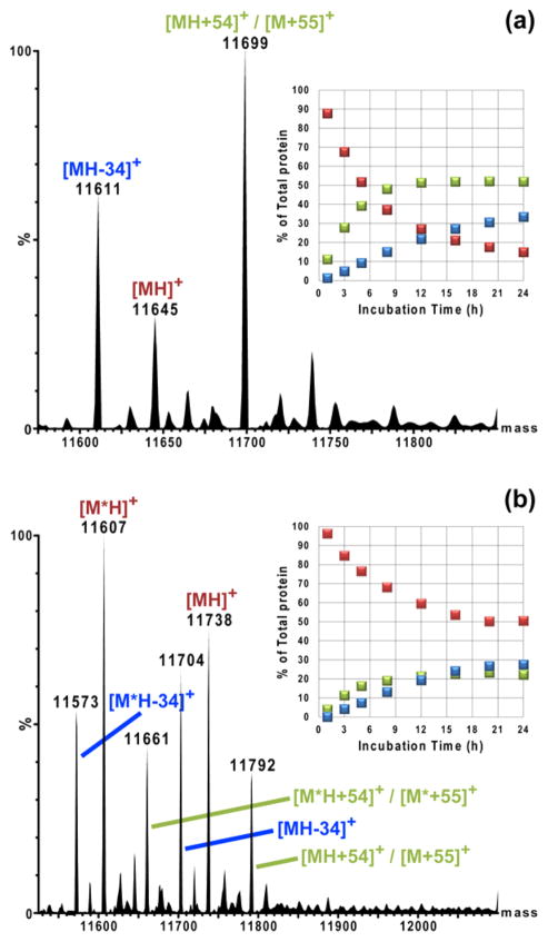
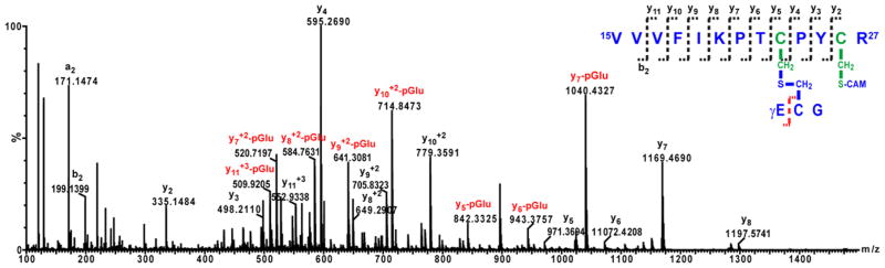
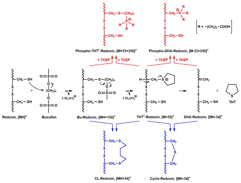
The myeloablative agent busulfan (1,4-butanediol dimethanesulfonate) is an old drug that is used routinely to eliminate cancerous bone marrow prior to hematopoietic stem cell transplant. The myeloablative activity and systemic toxicity of busulfan have been ascribed to its ability to cross-link DNA. In contrast, here we demonstrate that incubation of busulfan with the thiol redox proteins glutaredoxin or thioredoxin at pH 7.4 and 37 °C results in the formation of putative S-tetrahydrothiophenium adducts at their catalytic Cys residues, followed by β-elimination to yield dehydroalanine. Both proteins contain a second Cys, in their catalytic C-X-X-C motif, which reacts with the dehydroalanine, the initial Cys adduct with busulfan, or the S-tetrahydrothiophenium, to form novel intramolecular cross-links. The reactivity of the dehydroalanine (DHA) formed is further demonstrated by adduction with glutathione to yield a lanthionine and by a novel reaction with the reducing agent tris(2-carboxyethyl)phosphine (TCEP), which yields a phosphine adduct via Michael addition to the DHA. Formation of a second quaternary organophosphonium salt via nucleophilic substitution with TCEP on the initial busulfan-protein adduct or on the THT(+)-Redoxin species is also observed. These results reveal a rich potential for reactions of busulfan with proteins in vitro, and likely in vivo. It is striking that several of the chemically altered protein products retain none of the atoms of busulfan, in contrast to typical drug-protein adducts or traditional protein modification reagents. In particular, the ability of a clinically used drug to convert Cys to dehydrolanine in intact proteins, and its subsequent reaction with biological thiols, is unprecedented.
Conflict of interest statement
The authors declare no competing financial interest.
Figures


Similar articles
-
Electrophilic reactivity of the Busulfan metabolite, EdAG, towards cellular thiols and inhibition of human thioredoxin-1.Biochem Biophys Res Commun. 2020 Dec 10;533(3):325-331. doi: 10.1016/j.bbrc.2020.09.038. Epub 2020 Sep 18. Biochem Biophys Res Commun. 2020. PMID: 32958252
-
Characterization of alkali induced formation of lanthionine, trisulfides, and tetrasulfides from peptide disulfides using negative ion mass spectrometry.J Am Soc Mass Spectrom. 2009 May;20(5):783-91. doi: 10.1016/j.jasms.2008.12.019. Epub 2009 Jan 1. J Am Soc Mass Spectrom. 2009. PMID: 19200754
-
Glutathione conjugation of busulfan produces a hydroxyl radical-trapping dehydroalanine metabolite.Xenobiotica. 2012 Dec;42(12):1170-7. doi: 10.3109/00498254.2012.696740. Epub 2012 Jun 22. Xenobiotica. 2012. PMID: 22725664
-
High-dose treosulfan in conditioning prior to hematopoietic stem cell transplantation.Expert Opin Investig Drugs. 2010 Oct;19(10):1275-95. doi: 10.1517/13543784.2010.517744. Expert Opin Investig Drugs. 2010. PMID: 20836619 Review.
-
Chemistry, biochemistry, nutrition, and microbiology of lysinoalanine, lanthionine, and histidinoalanine in food and other proteins.J Agric Food Chem. 1999 Apr;47(4):1295-319. doi: 10.1021/jf981000+. J Agric Food Chem. 1999. PMID: 10563973 Review.
Cited by
-
The presence of busulfan metabolites and pharmacometabolomics in plasma drawn immediately before allograft infusion in hematopoietic cell transplant recipients.Clin Transl Sci. 2023 Dec;16(12):2577-2590. doi: 10.1111/cts.13651. Epub 2023 Oct 10. Clin Transl Sci. 2023. PMID: 37749994 Free PMC article.
-
Population pharmacokinetic model for once-daily intravenous busulfan in pediatric subjects describing time-associated clearance.CPT Pharmacometrics Syst Pharmacol. 2022 Aug;11(8):1002-1017. doi: 10.1002/psp4.12809. Epub 2022 Jun 16. CPT Pharmacometrics Syst Pharmacol. 2022. PMID: 35611997 Free PMC article.
-
Dynamics and inhibition of MLL1 CXXC domain on DNA revealed by single-molecule quantification.Biophys J. 2021 Aug 17;120(16):3283-3291. doi: 10.1016/j.bpj.2021.03.045. Epub 2021 Jul 17. Biophys J. 2021. PMID: 34280370 Free PMC article.
-
Review of the Pharmacokinetics and Pharmacodynamics of Intravenous Busulfan in Paediatric Patients.Clin Pharmacokinet. 2021 Jan;60(1):17-51. doi: 10.1007/s40262-020-00947-2. Epub 2020 Oct 30. Clin Pharmacokinet. 2021. PMID: 33128207 Review.
-
Characterization and quantitation of busulfan DNA adducts in the blood of patients receiving busulfan therapy.Mol Ther Oncolytics. 2023 Jan 20;28:197-210. doi: 10.1016/j.omto.2023.01.005. eCollection 2023 Mar 16. Mol Ther Oncolytics. 2023. PMID: 36820303 Free PMC article.
References
-
- McCune JS, Holmberg LA. Busulfan in hematopoietic stem cell transplant setting. Expert Opin Drug Metab Toxicol. 2009;5:957–969. - PubMed
-
- Ferry C, Socie G. Busulfan-cyclophosphamide versus total body irradiation-cyclophosphamide as preparative regimen before allogeneic hematopoietic stem cell transplantation for acute myeloid leukemia: what have we learned? Exp Hematol. 2003;31:1182–1186. - PubMed
-
- Barker CC, Butzner JD, Anderson RA, Brant R, Sauve RS. Incidence, survival and risk factors for the development of veno-occlusive disease in pediatric hematopoietic stem cell transplant recipients. Bone Marrow Transplant. 2003;32:79–87. - PubMed
-
- Reiss U, Cowan M, McMillan A, Horn B. Hepatic venoocclusive disease in blood and bone marrow transplantation in children and young adults: incidence, risk factors, and outcome in a cohort of 241 patients. J Pediatr Hematol/Oncol. 2002;24:746–750. - PubMed
Publication types
MeSH terms
Substances
Grants and funding
LinkOut - more resources
Full Text Sources
Other Literature Sources
