. 2017 Jan;32(1):60-69.
doi: 10.1002/jbmr.2934. Epub 2016 Sep 7.

Deletion of FoxO1, 3, and 4 in Osteoblast Progenitors Attenuates the Loss of Cancellous Bone Mass in a Mouse Model of Type 1 Diabetes

Affiliations

Deletion of FoxO1, 3, and 4 in Osteoblast Progenitors Attenuates the Loss of Cancellous Bone Mass in a Mouse Model of Type 1 Diabetes

Srividhya Iyer et al. J Bone Miner Res. 2017 Jan.

Abstract

Type 1 diabetes is associated with osteopenia and increased fragility fractures, attributed to reduced bone formation. However, the molecular mechanisms mediating these effects remain unknown. Insulin promotes osteoblast formation and inhibits the activity of the FoxO transcription factors. FoxOs, on the other hand, inhibit osteoprogenitor proliferation and bone formation. Here, we investigated whether FoxOs play a role in the low bone mass associated with type 1 diabetes, using mice lacking FoxO1, 3, and 4 in osteoprogenitor cells (FoxO1,3,4ΔOsx1-Cre ). Streptozotocin-induced diabetes caused a reduction in bone mass and strength in FoxO-intact mice. In contrast, cancellous bone was unaffected in diabetic FoxO1,3,4ΔOsx1-Cre mice. The low bone mass in the FoxO-intact diabetic mice was associated with decreased osteoblast number and bone formation, as well as decreased expression of the anti-osteoclastogenic cytokine osteoprotegerin (OPG) and increased osteoclast number. FoxO deficiency did not alter the effects of diabetes on bone formation; however, it did prevent the decrease in OPG and the increase in osteoclast number. Addition of high glucose to osteoblastic cell cultures decreased OPG mRNA, indicating that hyperglycemia in and of itself contributes to diabetic bone loss. Taken together, these results suggest that FoxOs exacerbate the loss of cancellous bone mass associated with type 1 diabetes and that inactivation of FoxOs might ameliorate the adverse effects of insulin deficiency. © 2016 American Society for Bone and Mineral Research.

Keywords: BONE HISTOMORPHOMETRY; DISEASES AND DISORDERS OF/RELATED TO BONE; GENETIC ANIMAL MODELS; STROMAL/STEM CELLS; TRANSCRIPTION FACTORS.


Conflict of interest statement

Disclosures

All authors state that they have no conflicts of interest.

Figures

Fig. 1
Fig. 1
Deletion of FoxOs in Osx1-Cre–expressing cells does not alter glucose metabolism. (A) Blood glucose of the 4-week-old male mice 7 days after being injected with vehicle or STZ for 5 consecutive days (n=8–15/group). (B) Body weight of the mice described in A, 40 days after the first injection. (C) Serum insulin levels in 3-month-old male mice at random feeding (n=8–10/group). Data presented as box plots with central box spanning 25th to 75th percentiles and the central line is mean. The whiskers represent the 10th and 90th percentiles, and values outside of this range are shown as dots.*p < 0.05 versus vehicle-treated mice of the same genotype; #p < 0.05 versus vehicle-treated control mice by two-way ANOVA. (D) Glucose tolerance test in 5 month old male mice (n=5–6/group). Data represent mean±SEM.
Fig. 2
Fig. 2
FoxO deletion in osteoblast progenitors prevents the loss of cancellous but not cortical bone in femora of diabetic mice. Micro-CT of femurs from mice injected with vehicle or STZ (n=8–15/group). (A) Cancellous bone volume and microarchitecture at the metaphysis. BV/TV=bone volume per tissue volume; Tb=trabecular. (B) Cortical thickness and bone area, total and medullary areas at the diaphysis.*p < 0.05 versus vehicle-treated mice of the same genotype; #p < 0.05 versus vehicle-treated control mice by two-way ANOVA.
Fig. 3
Fig. 3
The adverse effects of diabetes on cancellous bone are abrogated by FoxO deletion in osteoprogenitors. (A) Cancellous bone volume and microarchitecture of the lumbar vertebra (L5) from mice injected with vehicle or STZ (n=8–15/group). BV/TV=bone volume per tissue volume; Tb=trabecular. (B) Strength determined by compression (n=8–11/group). (C) Cortical thickness of the vertebral ventral wall.*p < 0.05 versus vehicle-treated mice of the same genotype; #p < 0.05 versus vehicle-treated control mice by two-way ANOVA.
Fig. 4
Fig. 4
Type 1 diabetes decreases bone formation independent of FoxOs in osteoblast lineage. (A) Osteoblast (Ob) number and surface per mm cancellous bone in vertebral bone sections from mice injected with vehicle or STZ (n=6/group). (B) Representative photomicrographs (scale bar=20 μm) and (C) mineralizing perimeter (M.Pm/B.Pm), mineral apposition (MAR), and bone formation rate (BFR) determined by tetracycline labels in sections described in A. (D) Gene expression determined by qRT-PCR in femoral and tibia shafts (n=8–15/group).*p < 0.05 versus vehicle-treated mice of same genotype and #p < 0.05 versus control mice with same treatment by two-way ANOVA.
Fig. 5
Fig. 5
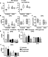
Type 1 diabetes decreases OPG and promotes bone resorption. (A) Osteoclast (Oc) number and perimeter quantified in vertebral sections (L1 to L3) from mice injected with vehicle or STZ (n=6/group). (B) mRNA by qRT-PCR of femur and tibia shafts (n=8–15/group).*p < 0.05 versus vehicle-treated mice of the same genotype; #p < 0.05 versus vehicle-treated control mice by two-way ANOVA. (C) Protein levels as determined by ELISA from bone marrow plasma of mice described in B. (D) OB-6 cells (triplicates) cultured with the indicated compounds for 48 hours; (E) calvaria cell cultures (triplicates). (F) Bone marrow stromal cells (triplicates) cultured in medium containing 1% ascorbate were treated with vehicle or 60 mM glucose.*p < 0.05 versus vehicle-treated mice of the same genotype; #p < 0.05 versus vehicle-treated control mice by two-way ANOVA. ≠p < 0.05 versus vehicle by one-way ANOVA.

Similar articles

Cited by

References

    1. Compston JE, Smith EM, Matthews C, Schofield P. Whole body composition and regional bone mass in women with insulin-dependent diabetes mellitus. Clin Endocrinol (Oxf) 1994;41(3):289–93. - PubMed
    1. Vestergaard P. Discrepancies in bone mineral density and fracture risk in patients with type 1 and type 2 diabetes—a meta-analysis. Osteoporos Int. 2007;18(4):427–44. - PubMed
    1. Hamann C, Kirschner S, Gunther KP, Hofbauer LC. Bone, sweet bone—osteoporotic fractures in diabetes mellitus. Nat Rev Endocrinol. 2012;8(5):297–305. - PubMed
    1. Khan TS, Fraser LA. Type 1 diabetes and osteoporosis: from molecular pathways to bone phenotype. J Osteoporos. 2015;2015:174186. - PMC - PubMed
    1. Hui SL, Epstein S, Johnston CC., Jr A prospective study of bone mass in patients with type I diabetes. J Clin Endocrinol Metab. 1985;60(1):74–80. - PubMed

Publication types

MeSH terms