Review
. 2017 Jan;101(1):25-30.
doi: 10.1136/bjophthalmol-2016-308823. Epub 2016 Aug 4.

Stargardt disease: clinical features, molecular genetics, animal models and therapeutic options

Affiliations
Review

Stargardt disease: clinical features, molecular genetics, animal models and therapeutic options

Preena Tanna et al. Br J Ophthalmol. 2017 Jan.

Abstract

Stargardt disease (STGD1; MIM 248200) is the most prevalent inherited macular dystrophy and is associated with disease-causing sequence variants in the gene ABCA4 Significant advances have been made over the last 10 years in our understanding of both the clinical and molecular features of STGD1, and also the underlying pathophysiology, which has culminated in ongoing and planned human clinical trials of novel therapies. The aims of this review are to describe the detailed phenotypic and genotypic characteristics of the disease, conventional and novel imaging findings, current knowledge of animal models and pathogenesis, and the multiple avenues of intervention being explored.

Keywords: Dystrophy; Imaging; Retina.


Conflict of interest statement

Conflicts of Interest: None declared.

Figures

Figure 1
Figure 1
(A) Colour fundus photograph showing typical yellow–white retinal flecks with macular atrophy. (B) Corresponding fundus autofluorescence image showing flecks of both increased and decreased autofluorescence and reduced central macular autofluorescence surrounded by an increased signal.
Figure 2
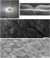
Figure 2
Multimodal imaging of the right eye of a molecularly proven 15-year-old patient with Stargardt disease. (A) Fundus autofluorescence image. The yellow line indicates the scanning level of the optical coherence tomography (OCT) scan in (B). (B) OCT scan showing central loss of outer retinal structure. The yellow arrows indicate the location and extent of the adaptive optics scanning light ophthalmoscopy (AOSLO) montages in (C) and (D) through the transition zone. (C) Confocal AOSLO montage of the photoreceptor mosaic. (D) Split-detection AOSLO montage of the photoreceptor mosaic. The far left side of (C) and (D) is closer to the fovea and lacks cone structure, corresponding with the lack of outer hyper-reflective layers in (B). Moving towards the right of (C) and (D), away from the fovea and superiorly on the retina, (C) shows structural changes that are relatively challenging to interpret, whereas (D) clearly shows the presence of cones. Scale bars represent 100 µm.

References

    1. Michaelides M, Hunt DM, Moore AT. The genetics of inherited macular dystrophies. J Med Genet 2003;40:641–50. 10.1136/jmg.40.9.641 - DOI - PMC - PubMed
    1. Fujinami K, Lois N, Davidson AE, et al. . A longitudinal study of stargardt disease: clinical and electrophysiologic assessment, progression, and genotype correlations. Am J Ophthalmol 2013;155:1075–88. 10.1016/j.ajo.2013.01.018 - DOI - PubMed
    1. Fujinami K, Zernant J, Chana RK, et al. . Clinical and molecular characteristics of childhood-onset Stargardt disease. Ophthalmology 2015;122:326–34. 10.1016/j.ophtha.2014.08.012 - DOI - PMC - PubMed
    1. Molday RS. Insights into the molecular properties of ABCA4 and its role in the visual cycle and stargardt disease. Prog Mol Biol Transl Sci 2015;134:415–31. 10.1016/bs.pmbts.2015.06.008 - DOI - PubMed
    1. Strauss RW, Ho A, Munoz B, et al. . The natural history of the progression of atrophy secondary to Stargardt disease (ProgStar) studies: design and baseline characteristics: ProgStar Report No. 1. Ophthalmology 2016;123:817–28. 10.1016/j.ophtha.2015.12.009 - DOI - PubMed

Substances