. 2016 Sep 2;4(9):726-33.
doi: 10.1158/2326-6066.CIR-16-0072. Epub 2016 Aug 4.

The Intratumoral Balance between Metabolic and Immunologic Gene Expression Is Associated with Anti-PD-1 Response in Patients with Renal Cell Carcinoma

Affiliations

The Intratumoral Balance between Metabolic and Immunologic Gene Expression Is Associated with Anti-PD-1 Response in Patients with Renal Cell Carcinoma

Maria Libera Ascierto et al. Cancer Immunol Res. .

Abstract

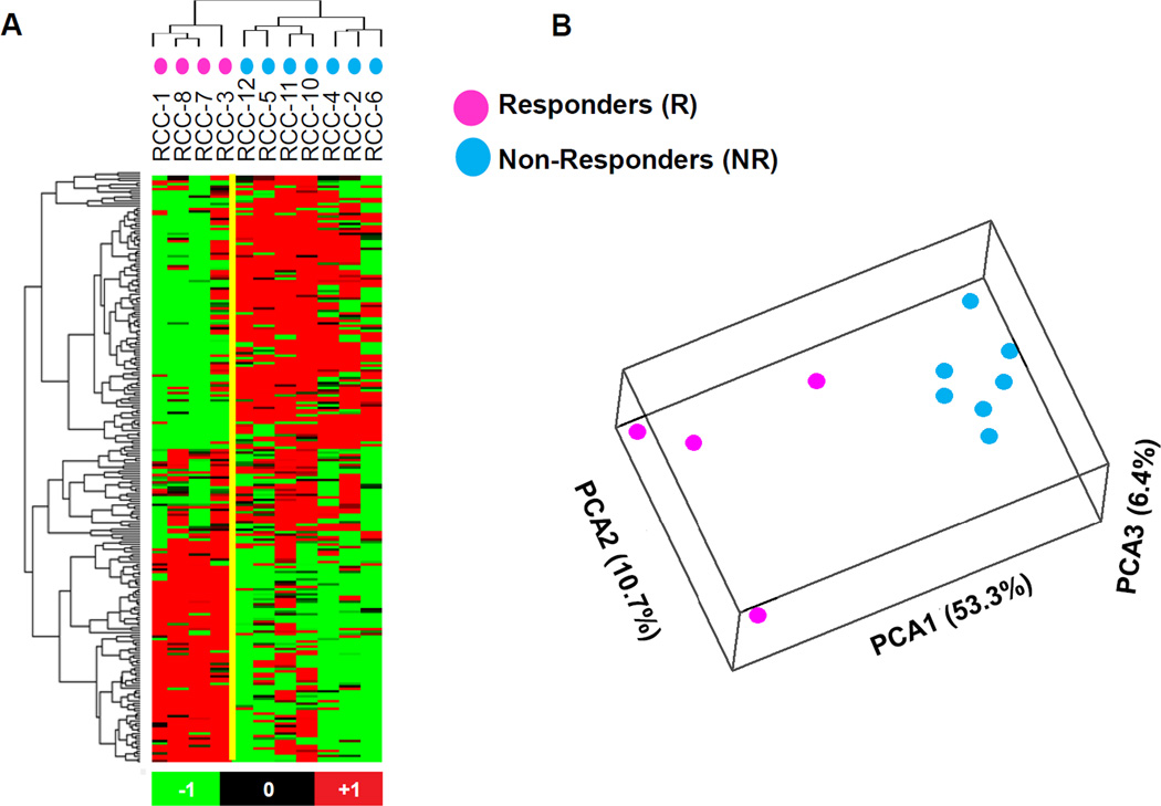
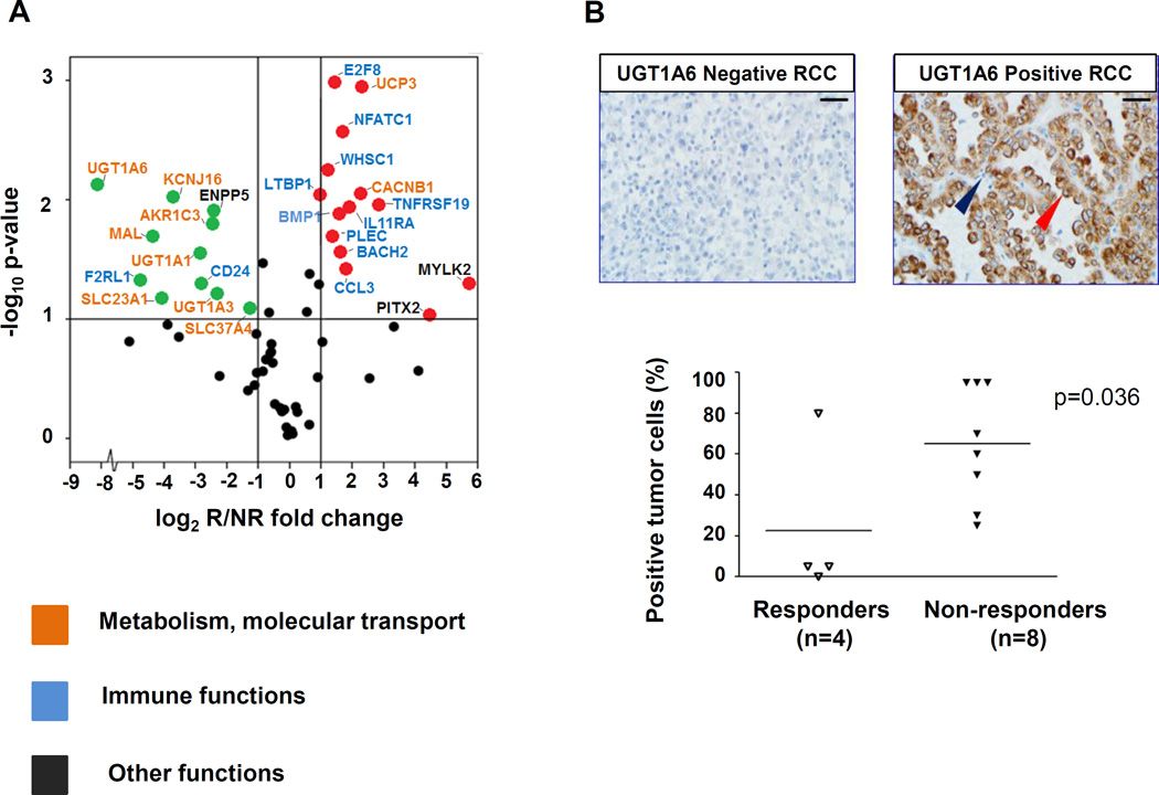
Pretreatment tumor PD-L1 expression has been shown to correlate with response to anti-PD-1/PD-L1 therapies. Yet, most patients with PD-L1(+) tumors do not respond to treatment. The current study was undertaken to investigate mechanisms underlying the failure of PD-1-targeted therapies in patients with advanced renal cell carcinoma (RCC) whose tumors express PD-L1. Formalin-fixed, paraffin-embedded pretreatment tumor biopsies expressing PD-L1 were derived from 13 RCC patients. RNA was isolated from PD-L1(+) regions and subjected to whole genome microarray and multiplex quantitative (q)RT-PCR gene expression analysis. A balance between gene expression profiles reflecting metabolic pathways and immune functions was associated with clinical outcomes following anti-PD-1 therapy. In particular, the expression of genes involved in metabolic and solute transport functions such as UGT1A family members, also found in kidney cancer cell lines, was associated with treatment failure in patients with PD-L1(+) RCC. Conversely, tumors from responding patients overexpressed immune markers such as BACH2, a regulator of CD4(+) T-cell differentiation, and CCL3 involved in leukocyte migration. These findings suggest that tumor cell-intrinsic metabolic factors may contribute to treatment resistance in RCC, thus serving as predictive markers for treatment outcomes and potential new targets for combination therapy regimens with anti-PD-1. Cancer Immunol Res; 4(9); 726-33. ©2016 AACRSee related Spotlight by Ohashi, p. 719.


Conflict of interest statement

The following authors have declared relevant financial relationships: MLA, TLM, AEB, LD, RAA, HX, TSP, JF, CC, and LC do not declare any conflicts.

Figures

Figure 1
Figure 1. Whole genome microarray analysis of pretreatment PD-L1 + RCC specimens demonstrates differential gene expression between patients responding or not to anti–PD-1 therapy
(A) Heat map and cluster analysis based on differentially expressed genes detected by 234 Illumina probes satisfying the criteria Student’s t test P ≤ 0.01 and fold change magnitude ≥ 1.5, comparing tumors from responders (R, n = 4) vs. nonresponders (NR, n = 7). Data were analyzed by using BRBArrayTools (http://linus.nci.nih.gov/BRB-ArrayTools.html) and visualized by Stanford Cluster Program and TreeView software. Red, high gene expression; green, low gene expression. (B) Principal component analysis (PCA) based on 1017 Illumina probes having differential expression in tumors from R vs. NR patients, with P ≤ 0.05 and fold change magnitude ≥1.5. Separation of the R and NR samples is seen. The principal component axis directions are labeled, with the percent of the total variance captured by each axis in parentheses.
Figure 2
Figure 2. Genes overexpressed in pretreatment PD-L1+ RCC specimens from responding vs. nonresponding patients reflect immune vs. metabolic functions, respectively
(A) Results of multiplex qRT-PCR for 60 select genes are shown, amplifying RNA isolated from pretreatment tumors in 4 responders and 8 nonresponders. Red and green dots represent genes significantly overexpressed or under-expressed, respectively, by at least 2-fold in tumors from responders compared to nonresponders, and with a 2-sided P value ≤ 0.1 (indicated by the horizontal line). Gene names are color-coded according to biological functions. The GUSB transcript was used as the internal reference. UGT1A6 was the most highly over-expressed gene associated with nonresponse to anti–PD-1. Similar results were obtained using 18S, ACTB, or PTPRC (CD45) as reference genes. Supporting information is provided in Table 2 and Supplementary Table S3. (B) UGT1A6 protein expression was evaluated by IHC in the same 12 pre-treatment PD-L1+ RCC specimens as were studied with qRT-PCR, including tumors from 4 responders and 8 nonresponders. In the top panels, representative UGT1A6 negative (left) and positive (right) RCC specimens are shown. Scale bars are equal to 25 um. Red arrow, kidney cancer cell with positive staining; blue arrow, infiltrating lymphocyte devoid of staining. In bottom panel, UGT1A6 expression is quantified by percent positive tumor cells in each specimen. Horizontal black bars indicate mean values. Enhanced UGT1A6 expression was significantly associated with nonresponse to anti–PD-1 therapy (P = 0.036, one-sided Wilcoxon rank sum test).

References

    1. Topalian SL, Hodi FS, Brahmer JR, Gettinger SN, Smith DC, McDermott DF, et al. Safety, activity, and immune correlates of anti-PD-1 antibody in cancer. N Engl J Med. 2012;366:2443–2454. - PMC - PubMed
    1. Brahmer JR, Tykodi SS, Chow LQ, Hwu WJ, Topalian SL, Hwu P, et al. Safety and activity of anti-PD-L1 antibody in patients with advanced cancer. N Engl J Med. 2012;366:2455–2465. - PMC - PubMed
    1. Brahmer JR, Drake CG, Wollner I, Powderly JD, Picus J, Sharfman WH, et al. Phase I study of single-agent anti-programmed death-1 (MDX-1106) in refractory solid tumors: safety, clinical activity, pharmacodynamics, and immunologic correlates. J Clin Oncol. 2010;28:3167–3175. - PMC - PubMed
    1. Hamid O, Robert C, Daud A, Hodi FS, Hwu WJ, Kefford R, et al. Safety and tumor responses with lambrolizumab (anti-PD-1) in melanoma. N Engl J Med. 2013;369:134–144. - PMC - PubMed
    1. Nghiem PT, Bhatia S, Lipson EJ, Kudchadkar RR, Miller NJ, Annamalai L, et al. PD-1 Blockade with Pembrolizumab in Advanced Merkel-Cell Carcinoma. N Engl J Med. 2016 [Epub ahead of print] - PMC - PubMed

Publication types

MeSH terms

Substances