. 2016 Aug 4;16(1):176.
doi: 10.1186/s12866-016-0793-5.

Plasmid pPCP1-derived sRNA HmsA promotes biofilm formation of Yersinia pestis

Affiliations

Plasmid pPCP1-derived sRNA HmsA promotes biofilm formation of Yersinia pestis

Zizhong Liu et al. BMC Microbiol. .

Abstract

Background: The ability of Yersinia pestis to form a biofilm is an important characteristic in flea transmission of this pathogen. Y. pestis laterally acquired two plasmids (pPCP1and pMT1) and the ability to form biofilms when it evolved from Yersinia pseudotuberculosis. Small regulatory RNAs (sRNAs) are thought to play a crucial role in the processes of biofilm formation and pathogenesis.

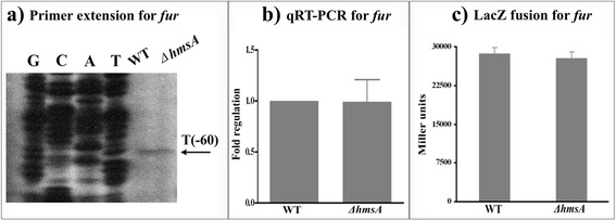
Results: A pPCP1-derived sRNA HmsA (also known as sR084) was found to contribute to the enhanced biofilm formation phenotype of Y. pestis. The concentration of c-di-GMP was significantly reduced upon deletion of the hmsA gene in Y. pestis. The abundance of mRNA transcripts determining exopolysaccharide production, crucial for biofilm formation, was measured by primer extension, RT-PCR and lacZ transcriptional fusion assays in the wild-type and hmsA mutant strains. HmsA positively regulated biofilm synthesis-associated genes (hmsHFRS, hmsT and hmsCDE), but had no regulatory effect on the biofilm degradation-associated gene hmsP. Interestingly, the recently identified biofilm activator sRNA, HmsB, was rapidly degraded in the hmsA deletion mutant. Two genes (rovM and rovA) functioning as biofilm regulators were also found to be regulated by HmsA, whose regulatory effects were consistent with the HmsA-mediated biofilm phenotype.

Conclusion: HmsA potentially functions as an activator of biofilm formation in Y. pestis, implying that sRNAs encoded on the laterally acquired plasmids might be involved in the chromosome-based regulatory networks implicated in Y. pestis-specific physiological processes.


Figures

Fig. 1
Fig. 1
Biofilm formation capacity and c-di-GMP production mediated by HmsA. a Bacterial colony morphology. The WT::pBAD, ∆hmsA::pBAD and ∆hmsA::HmsA strains were incubated on LB agar with 0.1 % arabinose designated as “+Ara” (middle row). The ∆hmsS or ∆fur strains were used as negative or positive controls for this assay, respectively (bottom row). b Crystal violet staining. Biofilms were quantified by crystal violet staining in the WT, ∆hmsA and ∆hmsA::HmsA strains grown in LB medium with the addition of 0.1 % arabinose. The ∆hmsS strain was grown under the same conditions as the negative control. Data are presented as the average of three separate experiments and error bars represent the standard deviation. c C. elegans biofilms. The percentage of L4/adults after incubation of nematode eggs on a lawn of the indicated Y. pestis strains was used to evaluate the capacity for biofilm formation. d Intracellular c-di-GMP concentration
Fig. 2
Fig. 2
Regulatory effects of HmsA on hmsH. a Primer extension results. The relative levels of hmsH transcript were determined in the WT and ∆hmsA mutant strains by primer extension assays. The Sanger sequence ladders (lanes G, C, A and T) and the primer extension products of hmsH were analyzed on an 8 M urea-6 % acrylamide sequencing gel. The transcription start sites of hmsH were indicated by arrows, and the minus number under the arrow indicates the nucleotide position upstream of the hmsH start codon. b qRT-PCR results. The relative levels of the hmsH transcript were determined in the WT and ∆hmsA mutant strains by qRT-PCR. c lacZ fusion results. The hmsH::lacZ transcriptional fusion vector was transformed into the WT and ∆hmsA mutant strains. The transcriptional activity of hmsH determined in the bacterial cellular extracts was represented as Miller units of β-galactosidase activity
Fig. 3
Fig. 3
Regulatory effects of HmsA on hmsT. a Primer extension results. The relative levels of hmsH transcript were determined in the WT and ∆hmsA mutant strains by primer extension assays. The Sanger sequence ladders (lanes G, C, A and T) and the primer extension products of hmsH were analyzed on an 8 M urea-6 % acrylamide sequencing gel. The transcription start sites of hmsH were indicated by arrows, and the minus number under the arrow indicates the nucleotide position upstream of the hmsH start codon. b qRT-PCR results. The relative levels of the hmsH transcript were determined in the WT and ∆hmsA mutant strains by qRT-PCR. c lacZ fusion results. The hmsH::lacZ transcriptional fusion vector was transformed into the WT and ∆hmsA mutant strains. The transcriptional activity of hmsH determined in the bacterial cellular extracts was represented as Miller units of β-galactosidase activity
Fig. 4
Fig. 4
Regulatory effects of HmsA on hmsC. a Primer extension results. The relative levels of hmsH transcript were determined in the WT and ∆hmsA mutant strains by primer extension assays. The Sanger sequence ladders (lanes G, C, A and T) and the primer extension products of hmsH were analyzed on an 8 M urea-6 % acrylamide sequencing gel. The transcription start sites of hmsH were indicated by arrows, and the minus number under the arrow indicates the nucleotide position upstream of the hmsH start codon. b qRT-PCR results. The relative levels of the hmsH transcript were determined in the WT and ∆hmsA mutant strains by qRT-PCR. c lacZ fusion results. The hmsH::lacZ transcriptional fusion vector was transformed into the WT and ∆hmsA mutant strains. The transcriptional activity of hmsH determined in the bacterial cellular extracts was represented as Miller units of β-galactosidase activity
Fig. 5
Fig. 5
Regulatory effects of HmsA on hmsP. a Primer extension results. The relative levels of hmsH transcript were determined in the WT and ∆hmsA mutant strains by primer extension assays. The Sanger sequence ladders (lanes G, C, A and T) and the primer extension products of hmsH were analyzed on an 8 M urea-6 % acrylamide sequencing gel. The transcription start sites of hmsH were indicated by arrows, and the minus number under the arrow indicates the nucleotide position upstream of the hmsH start codon. b qRT-PCR results. The relative levels of the hmsH transcript were determined in the WT and ∆hmsA mutant strains by qRT-PCR. c lacZ fusion results. The hmsH::lacZ transcriptional fusion vector was transformed into the WT and ∆hmsA mutant strains. The transcriptional activity of hmsH determined in the bacterial cellular extracts was represented as Miller units of β-galactosidase activity
Fig. 6
Fig. 6
Regulatory effects of HmsA on HmsB. a Primer extension results. b Confirmation of HmsA and HmsB by northern blot analysis. The transcript of hmsB/hmsA in the WT and ∆hmsA/hmsB mutant strains was monitored by northern blotting (upper and bottom panel). c Measurement of the half-life of HmsB. The WT and ∆hmsA strains were grown to exponential phase at 26 °C and then treated with 250 μg/mL of rifampicin. Culture samples were collected at 0, 2, 4, 8, 16 and 32 min and were subject to RNA extraction and northern blotting using 5S rRNA and HmsB probes
Fig. 7
Fig. 7
Regulatory effects of HmsA on rovA. a Primer extension results. The relative levels of hmsH transcript were determined in the WT and ∆hmsA mutant strains by primer extension assays. The Sanger sequence ladders (lanes G, C, A and T) and the primer extension products of hmsH were analyzed on an 8 M urea-6 % acrylamide sequencing gel. The transcription start sites of hmsH were indicated by arrows, and the minus number under the arrow indicates the nucleotide position upstream of the hmsH start codon. b qRT-PCR results. The relative levels of the hmsH transcript were determined in the WT and ∆hmsA mutant strains by qRT-PCR. c lacZ fusion results. The hmsH::lacZ transcriptional fusion vector was transformed into the WT and ∆hmsA mutant strains. The transcriptional activity of hmsH determined in the bacterial cellular extracts was represented as Miller units of β-galactosidase activity
Fig. 8
Fig. 8
Regulatory effects of HmsA on rovM. a Primer extension results. The relative levels of hmsH transcript were determined in the WT and ∆hmsA mutant strains by primer extension assays. The Sanger sequence ladders (lanes G, C, A and T) and the primer extension products of hmsH were analyzed on an 8 M urea-6 % acrylamide sequencing gel. The transcription start sites of hmsH were indicated by arrows, and the minus number under the arrow indicates the nucleotide position upstream of the hmsH start codon. b qRT-PCR results. The relative levels of the hmsH transcript were determined in the WT and ∆hmsA mutant strains by qRT-PCR. c lacZ fusion results. The hmsH::lacZ transcriptional fusion vector was transformed into the WT and ∆hmsA mutant strains. The transcriptional activity of hmsH determined in the bacterial cellular extracts was represented as Miller units of β-galactosidase activity
Fig. 9
Fig. 9
Regulatory effects of HmsA on fur. a Primer extension results. The relative levels of hmsH transcript were determined in the WT and ∆hmsA mutant strains by primer extension assays. The Sanger sequence ladders (lanes G, C, A and T) and the primer extension products of hmsH were analyzed on an 8 M urea-6 % acrylamide sequencing gel. The transcription start sites of hmsH were indicated by arrows, and the minus number under the arrow indicates the nucleotide position upstream of the hmsH start codon. b qRT-PCR results. The relative levels of the hmsH transcript were determined in the WT and ∆hmsA mutant strains by qRT-PCR. c lacZ fusion results. The hmsH::lacZ transcriptional fusion vector was transformed into the WT and ∆hmsA mutant strains. The transcriptional activity of hmsH determined in the bacterial cellular extracts was represented as Miller units of β-galactosidase activity
Fig. 10
Fig. 10
Regulatory networks of HmsA on biofilm formation

Similar articles

Cited by

References

    1. Rasmussen S, Allentoft ME, Nielsen K, Orlando L, Sikora M, Sjogren KG, Pedersen AG, Schubert M, Van Dam A, Kapel CM, et al. Early divergent strains of Yersinia pestis in Eurasia 5,000 years ago. Cell. 2015;163(3):571–82. doi: 10.1016/j.cell.2015.10.009. - DOI - PMC - PubMed
    1. Erickson DL, Jarrett CO, Wren BW, Hinnebusch BJ. Serotype differences and lack of biofilm formation characterize Yersinia pseudotuberculosis infection of the Xenopsylla cheopis flea vector of Yersinia pestis. J Bacteriol. 2006;188(3):1113–9. doi: 10.1128/JB.188.3.1113-1119.2006. - DOI - PMC - PubMed
    1. Zhou D, Yang R. Formation and regulation of Yersinia biofilms. Protein Cell. 2011;2(3):173–9. doi: 10.1007/s13238-011-1024-3. - DOI - PMC - PubMed
    1. Perry RD, Fetherston JD. Yersinia pestis--etiologic agent of plague. Clin Microbiol Rev. 1997;10(1):35–66. - PMC - PubMed
    1. Ramos JL, Gallegos MT, Marques S, Ramos-Gonzalez MI, Espinosa-Urgel M, Segura A. Responses of Gram-negative bacteria to certain environmental stressors. Curr Opin Microbiol. 2001;4(2):166–71. doi: 10.1016/S1369-5274(00)00183-1. - DOI - PubMed

MeSH terms

LinkOut - more resources