. 2016 Dec 5:437:108-119.
doi: 10.1016/j.mce.2016.07.040. Epub 2016 Aug 1.

Agonist-induced internalization and desensitization of the apelin receptor

Affiliations

Agonist-induced internalization and desensitization of the apelin receptor

George R Pope et al. Mol Cell Endocrinol. .

Abstract

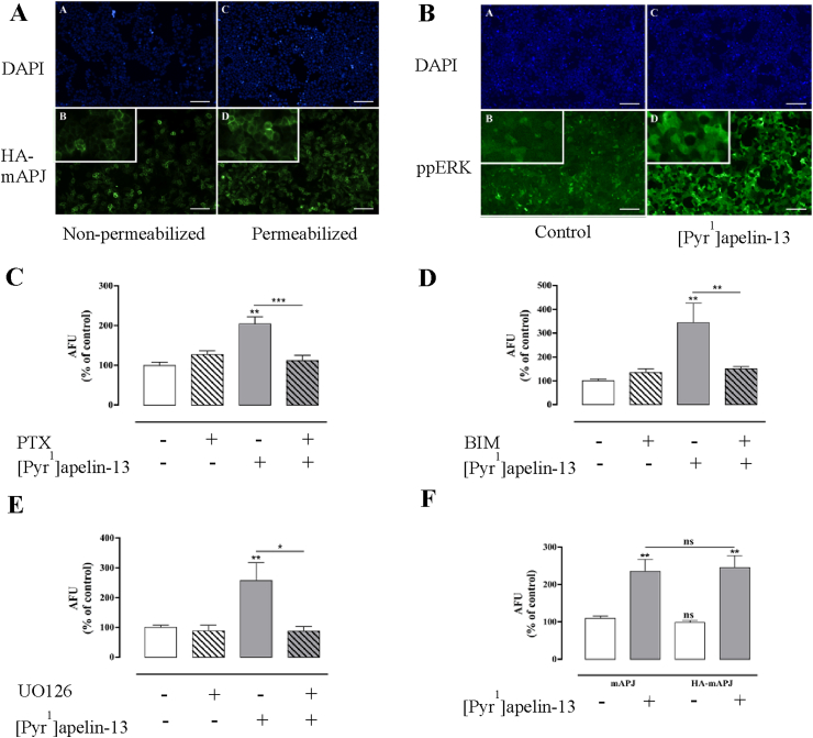
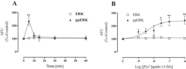
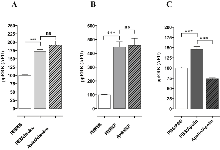
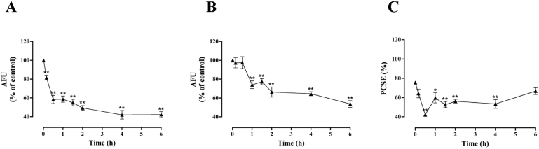
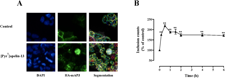
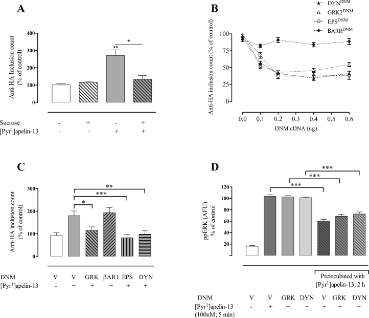
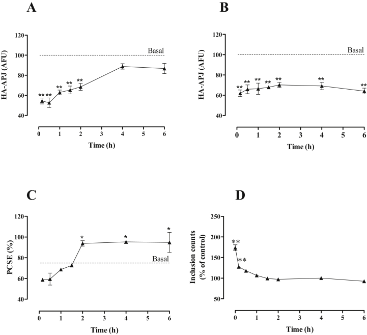
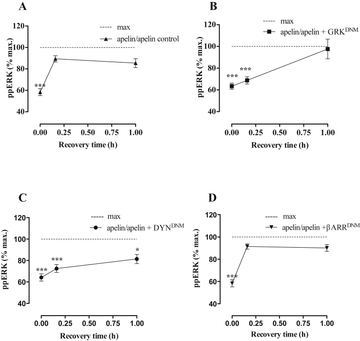
Apelin acts via the G protein-coupled apelin receptor (APJ) to mediate effects on cardiovascular and fluid homeostasis. G protein-coupled receptor (GPCR) trafficking has an important role in the regulation of receptor signalling pathways and cellular functions, however in the case of APJ the mechanisms and proteins involved in apelin-induced trafficking are not well understood. We generated a stable HEK-293 cell line expressing N-terminus HA-tagged mouse (m) APJ, and used a semi-automated imaging protocol to quantitate APJ trafficking and ERK1/2 activation following stimulation with [Pyr1]apelin-13. The mechanisms of [Pyr1]apelin-13-induced internalization and desensitization were explored using dominant-negative mutant (DNM) cDNA constructs of G protein-coupled receptor kinase 2 (GRK2), β-arrestin1, EPS15 and dynamin. The di-phosphorylated ERK1/2 (ppERK1/2) response to [Pyr1]apelin-13 desensitized during sustained stimulation, due to upstream APJ-specific adaptive changes. Furthermore, [Pyr1]apelin-13 stimulation caused internalization of mAPJ via clathrin coated vesicles (CCVs) and also caused a rapid reduction in cell surface and whole cell HA-mAPJ. Our data suggest that upon continuous agonist exposure GRK2-mediated phosphorylation targets APJ to CCVs that are internalized from the cell surface in a β-arrestin1-independent, EPS15- and dynamin-dependent manner. Internalization does not appear to contribute to the desensitization of APJ-mediated ppERK1/2 activation in these cells.

Keywords: Apelin; Apelin receptor; Extracellular-signal-regulated kinase (ERK); G protein-coupled receptor; Intracellular trafficking; Signalling.


Figures

Fig. 1
Fig. 1
(A) Thumbnail images from individual wells stained for HA-mAPJ cell surface and whole cell expression, after stimulation with [Pyr1]apelin-13. Representative regions of cell images are shown for DAPI (top panels A and C), and HA-mAPJ (bottom panels B and D) in HA-mAPJ-HEK293 cells with either non-permeabilized (left panels, cell surface) or permeabilized (right panels, whole cell) membranes, higher magnification inset. (B) Thumbnail images from individual wells stained for ppERK1/2 expression after stimulation with [Pyr1]apelin-13. Representative regions of cell images are shown for DAPI (top panels A and C), and ppERK1/2 (bottom panels B and D) in mAPJ-HEK293 cells stimulated with either vehicle control (left panels) or 100 nM [Pyr1]apelin-13 (right panels), higher magnification inset. Scale bars, 100 μm. mAPJ-HEK293 cells were pre-treated with or without (C) PTX (200 ng/ml, 16 h), (D) BIM (10 μM, 1 h) or (E) UO126 (10 μM, 30 min) and stimulated in the presence or absence of [Pyr1]apelin-13 (100 nM) for 5 min. (F) Tagging of mAPJ with the HA epitope did not interfere with receptor signalling. Non tagged mAPJ-HEK293 cells and HA-tagged mAPJ-HEK293 cells were stimulated with [Pyr1]apelin-13 (100 nM) for 5 min and compared with control cells treated with 1× PBS. For (CF) cells were fixed, stained, and imaged for determination of whole-cell ppERK1/2 intensity using anti ppERK1/2 antibody. The value determined with no primary antibody present was designated as background and was subtracted from raw data to give arbitrary fluorescence units (AFU) and then normalized to a percentage of vehicle control. Data shown are mean ± SEM, of at least three separate experiments, each with triplicate wells and triplicate fields within wells. *p < 0.05, **p < 0.01, and ***p < 0.001 comparing stimulations to basal conditions, analysed by one-way ANOVA and Dunnett's multiple comparison post hoc tests. ns = no statistical significant difference.
Fig. 2
Fig. 2
(A) [Pyr1]apelin-13 stimulated ERK activation in mAPJ-HEK293 at the time points indicated and (B) with the indicated concentrations of [Pyr1]apelin-13 for 5 min. Cells were fixed, stained, and imaged for determination of whole-cell ppERK1/2 using anti ppERK1/2 antibody, and total ERK1/2 measured with anti-ERK1/2 antibody. The value determined with no primary antibody present was designated as background and was subtracted from raw data to give arbitrary fluorescence units (AFU) and then normalized to a percentage of vehicle control. Data shown are mean ± SEM, of at least three separate experiments, each with triplicate wells and triplicate fields within wells. *p < 0.05 and **p < 0.01 comparing stimulations to basal conditions, analysed by one-way ANOVA and Dunnett's multiple comparison post hoc tests.
Fig. 3
Fig. 3
Desensitization of [Pyr1]apelin-13-induced ERK1/2 activation in mAPJ-HEK293. mAPJ-HEK293 cells were pre-incubated with PBS or [Pyr1]apelin-13 (100 nM, 2 h), washed, and immediately stimulated in the presence of (A) adrenaline (1 μM) (B) EGF (100 ng/ml) or (C) [Pyr1]apelin-13 (100 nM) for 5 min. Cells were fixed, stained and imaged for determination of whole cell ppERK1/2 intensity using anti-ppERK1/2 antibody. The value determined with no primary antibody present was designated as background and was subtracted from raw data to give ppERK1/2 intensity in arbitrary fluorescence units (AFU) and then normalized to a percentage of vehicle control. Data shown are mean ± SEM, of at least three separate experiments, each with triplicate wells and triplicate fields within wells. ***p < 0.001, analysed by one-way ANOVA and Dunnett’s multiple comparison post hoc tests. ns = no statistical significant difference.
Fig. 4
Fig. 4
Time course of [Pyr1]apelin-13-induced HA-mAPJ localization and expression levels. HA-mAPJ-HEK293 cells were incubated in the presence or absence of [Pyr1]apelin-13 (100 nM) for 0–6 h. Cells were fixed, stained and imaged for determination of either (A) cell surface or (B) whole cell HA-mAPJ intensity using anti-HA antibody. The value determined with no primary antibody present was designated as background and was subtracted from raw data to give HA-mAPJ intensity in arbitrary fluorescence units (AFU) and then normalized to a percentage of vehicle control. Cell surface and whole cell AFU values were used to determine the PCSE (C). Data shown are mean ± SEM, of at least three separate experiments, each with triplicate wells and triplicate fields within wells. * = p < 0.05 and ** = p < 0.01, comparing stimulations to basal conditions, analysed by one-way ANOVA and Dunnett's multiple comparison post hoc tests.
Fig. 5
Fig. 5
(A) Representative regions of cell images shown for DAPI, HA-mAPJ and an illustration of the automated image segmentation used to define perimeters of nuclei (blue) and cells (green or red) and inclusions (yellow) in cells stimulated with control or [Pyr1]apelin-13, as indicated. (B) Time course of [Pyr1]apelin-13–induced HA-mAPJ internalization. HA-mAPJ-HEK293 cells were pre-treated with anti-HA antibody (1:1000; 1 h 37 °C/5% CO2), washed, then incubated in the presence or absence of [Pyr1]apelin-13 (100 nM) for 0–6 h. Data shown are mean ± SEM, of at least three separate experiments, each with triplicate wells and triplicate fields within wells. **p < 0.01, comparing stimulations to basal conditions, analysed by one-way ANOVA and Dunnett's multiple comparison post hoc tests. (For interpretation of the references to colour in this figure legend, the reader is referred to the web version of this article.)
Fig. 6
Fig. 6
Mechanisms of [Pyr1]apelin-13-induced HA-mAPJ internalization and mAPJ desensitization. (A) HA-mAPJ-HEK293 cells were pre-treated with anti-HA antibody (1:100; 1 h, 37 °C/5% CO2), washed, then incubated in the presence or absence of sucrose (0.4 M) for 45 min, followed by incubation with [Pyr1]apelin-13 (100 nM) for 2 h. (B, C) HA-mAPJ-HEK293 cells were transfected with DNM cDNAs of various CME-related factors or an empty plasmid vector (V). After 48 h, cells were pre-treated with anti-HA antibody (1:100; 1 h, 37 °C/5% CO2), washed, incubated in the presence or absence of [Pyr1]apelin-13 (100 nM), for 2 h. Cells were fixed, stained and imaged for determination of whole cell inclusion counts, normalized to a percentage of vehicle control. (B) shows [Pyr1]apelin-13-induced HA-mAPJ internalization after co-transfection with increasing concentrations of GRKDNM, EPSDNM, DYNDNM and βARRDNM. (C) shows the effects of GRKDNM, EPSDNM, DYNDNM and βARRDNM on [Pyr1]apelin-13-induced HA-mAPJ internalization. In (D) mAPJ-HEK293 cells were transfected with DNM cDNAs of various CME-related factors or an empty plasmid vector (V). After 48 h, cells were washed, incubated in the presence or absence of [Pyr1]apelin-13 (100 nM), for 2 h, stimulated with 100 nM [Pyr1]apelin-13 for 5 min, fixed, stained and imaged for determination of whole cell ppERK1/2 intensity using anti-ppERK1/2 antibody, expressed as arbitrary fluorescent units (AFU). Data shown are mean ± SEM, of at least three separate experiments, each with triplicate wells and triplicate fields within wells. *p < 0.05, **p < 0.01, ***p < 0.001 analysed by two-way ANOVA and Dunnett's multiple comparison post hoc tests.
Fig. 7
Fig. 7
Recovery of APJ levels after agonist removal. HA-mAPJ-HEK293 cells were incubated in the presence or absence of [Pyr1]apelin-13 (100 nM) for 2 h, washed, then incubated in fresh medium for 0–6 h. Cells were fixed, stained and imaged for determination of either (A) cell surface or (B) whole cell HA-mAPJ intensity using anti-HA antibody. The value determined with no primary antibody present was designated as background and was subtracted from raw data to give HA-mAPJ intensity in arbitrary fluorescence units (AFU) and then normalized to a percentage of vehicle control. Cell surface and whole cell AFU values were used to determine the PCSE (C). (D) shows recovery from the effect of [Pyr1]apelin-13 on HA-mAPJ inclusion count. HA-mAPJ-HEK293 cells were pre-treated with anti-HA antibody (1:100; 1 h, 37 °C/5% CO2), washed, incubated in the presence or absence of [Pyr1]apelin-13 (100 nM) for 2 h to internalize APJ, washed, then incubated in fresh medium for 0–6 h. Data shown are mean ± SEM, of three separate experiments, each with triplicate wells and triplicate fields within wells. *p < 0.05 and **p < 0.01, comparing stimulations to basal conditions, analysed by one-way ANOVA and Dunnett's multiple comparison post hoc tests.
Fig. 8
Fig. 8
(A) mAPJ-HEK293 cells were pre-incubated in the presence or absence of [Pyr1]apelin-13 (100 nM, 2 h), washed, incubated in fresh medium for 0–1 h and then stimulated in the presence or absence of [Pyr1]apelin-13 (100 nM) for 5 min. Cells were fixed, stained and imaged for determination of whole cell ppERK1/2 intensity using anti-ppERK1/2 antibody. The value determined with no primary antibody present was designated as background and was subtracted from raw data to give ppERK1/2 intensity in arbitrary fluorescence units (AFU) and then normalized to a percentage of vehicle control ([Pyr1]apelin-13-induced ERK1/2 signalling in cells initially exposed to vehicle control and designated as “max”). In (BD) mAPJ HEK293 cell lines were transfected with GRKDNM, DYNDNM or βARRDNM cDNAs respectively before preincubation with or without [Pyr1]apelin-13. Data shown are mean ± SEM, of at least three separate experiments, each with triplicate wells and triplicate fields within wells. ***p < 0.001, comparing stimulations to max conditions, analysed by one-way ANOVA and Dunnett's multiple comparison post hoc tests.

References

    1. Ahn S., Shenoy S.K., Wei H., Lefkowitz R.J. Differential kinetic and spatial patterns of beta-arrestin and G protein-mediated ERK activation by the angiotensin II receptor. J. Biol. Chem. 2004;279:35518–35525. - PubMed
    1. Bai B., Tang J., Liu H., Chen J., Li Y., Song W. Apelin-13 induces ERK1/2 but not p38 MAPK activation through coupling of the human apelin receptor to the Gi2 pathway. Acta Biochim. Biophys. Sin. (Shanghai) 2008;40:311–318. - PubMed
    1. Benovic J.L., Pike L.J., Cerione R.A., Staniszewski C., Yoshimasa T., Codina J., Caron M.G., Lefkowitz R.J. Phosphorylation of the mammalian beta-adrenergic receptor by cyclic AMP dependent protein kinase. Regulation of the rate of receptor phosphorylation and dephosphorylation by agonist occupancy and effects on coupling of the receptor to the stimulatory guanine nucleotide regulatory protein. J. Biol. Chem. 1985;260:7094–7101. - PubMed
    1. Benovic J.L., Strasser R.H., Caron M.G., Lefkowitz R.J. Beta-adrenergic receptor kinase: identification of a novel protein kinase that phosphorylates the agonist-occupied form of the receptor. Proc. Natl. Acad. Sci. U. S. A. 1986;83:2797–2801. - PMC - PubMed
    1. Bhatnagar A., Willins D.L., Gray J.A., Woods J., Benovic J.L., Roth B.L. The dynamin-dependent, arrestin-independent internalization of 5-hydroxytryptamine 2A (5-HT2A) serotonin receptors reveals differential sorting of arrestins and 5-HT2A receptors during endocytosis. J. Biol. Chem. 2001;276:8269–8277. - PubMed

Publication types

MeSH terms

Substances

LinkOut - more resources