. 2016 Aug 5:6:30282.
doi: 10.1038/srep30282.

Lipid Regulated Intramolecular Conformational Dynamics of SNARE-Protein Ykt6

Affiliations

Lipid Regulated Intramolecular Conformational Dynamics of SNARE-Protein Ykt6

Yawei Dai et al. Sci Rep. .

Abstract

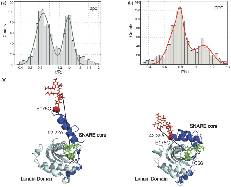
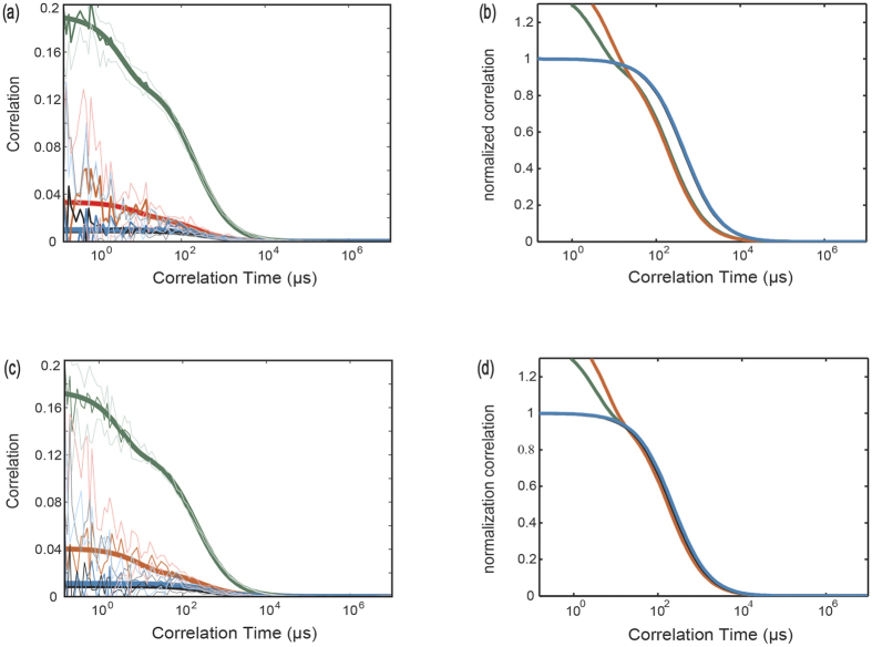
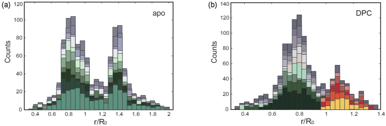
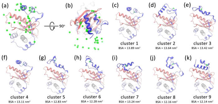
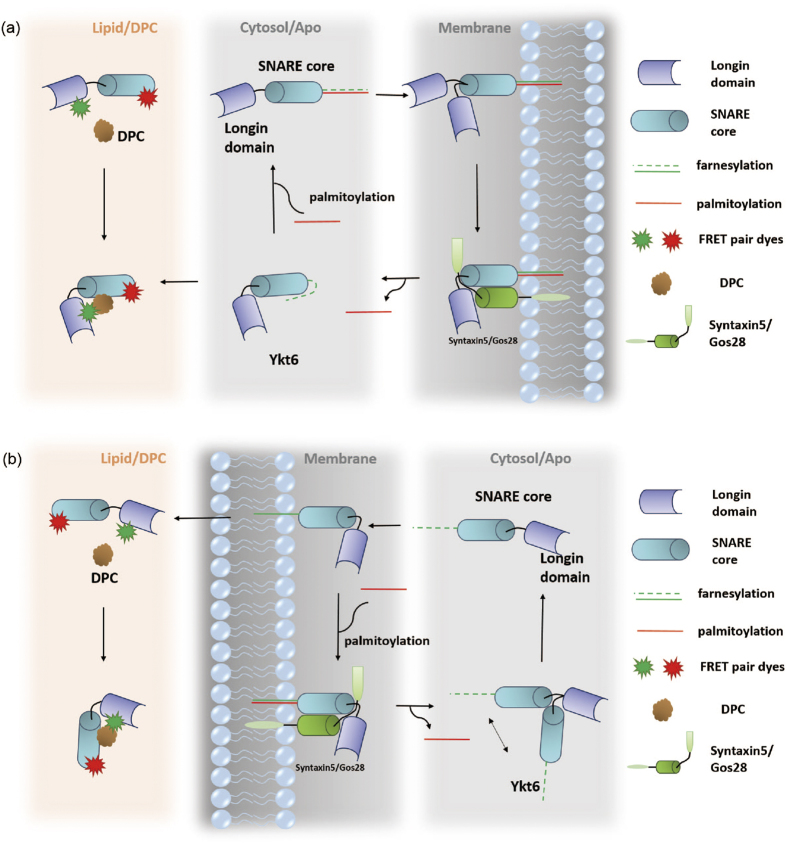
Cellular informational and metabolic processes are propagated with specific membrane fusions governed by soluble N-ethylmaleimide sensitive factor attachment protein receptors (SNARE). SNARE protein Ykt6 is highly expressed in brain neurons and plays a critical role in the membrane-trafficking process. Studies suggested that Ykt6 undergoes a conformational change at the interface between its longin domain and the SNARE core. In this work, we study the conformational state distributions and dynamics of rat Ykt6 by means of single-molecule Förster Resonance Energy Transfer (smFRET) and Fluorescence Cross-Correlation Spectroscopy (FCCS). We observed that intramolecular conformational dynamics between longin domain and SNARE core occurred at the timescale ~200 μs. Furthermore, this dynamics can be regulated and even eliminated by the presence of lipid dodecylphoshpocholine (DPC). Our molecular dynamic (MD) simulations have shown that, the SNARE core exhibits a flexible structure while the longin domain retains relatively stable in apo state. Combining single molecule experiments and theoretical MD simulations, we are the first to provide a quantitative dynamics of Ykt6 and explain the functional conformational change from a qualitative point of view.


Figures

Figure 1
Figure 1. SmFRET Distance Distributions and structures of Ykt6 in the apo and with DPC conditions.
(a) Apo condition: Distribution of SNARE core to longin domain distances from smFRET results for doubly labeled Ykt6 molecules before adding DPC. Fitting results with two Gaussians are plotted in green (Peaks are centred at 0.85R0 and 1.39R0, respectively). (b) DPC condition: Distributions of distance from smFRET results for double labeled Ykt6 molecules in the presence of DPC environment (Ykt6:DPC = 1:10). The Gaussian fitting lines also are included in red line (Peaks are centre at 0.82R0 and 1.09R0, respectively). (c) Two structures predicted by our MD simulations showing the structures of doubly labeled Ykt6 in open (left) and closed (right) states. Both structures were simulated based on PDB structure 3KYQ, which was closer to the closed form. Structures of dyes were simulated with assumed structures of Alexa 555 (green) and Alexa 647 (red).
Figure 2
Figure 2. FCCS data for apo and saturated DPC (Ykt6:DPC = 1:2) conditions (green: AC-d; red: AC-a; black: CC-da; blue: CC-ad).
(a) Raw FCCS time correlation function data of rYkt6ΔC (all FCCS experiments are done on samples labeled with Alexa 488/647 FRET pairs) in the apo environment free of DPC. The thin fluctuated lines are the original data, the bold coloured curves are smoothed by increasing the number of binsize in triplet, whereas the thick lines indicate the fitted curves. (b) Normalized FCCS data of rYkt6ΔC in the apo environment free of DPC. (c) Raw FCCS data of rYkt6ΔC in the environment of Ykt6:DPC = 1:2. (d) Normalized FCCS data of rYkt6ΔC in the environment Ykt6:DPC = 1:2.
Figure 3
Figure 3. Distance distributions of single trajectory of Ykt6 with/without DPC.
(a) In the apo condition, we plotted the histogram of Ykt6 SNARE core to longin distances from the smFRET experiment. The whole histogram was divided into different colour blocks. Each colour represents the distance statistics from the same trajectory, and therefore the same molecule. From this plot we can see, each molecule monitored in our apo smFRET experiment contributes to both open and closed states during the observation time. (b) When the same plot is generated for the DPC environment (Ykt6:DPC = 1:10), the data from each molecule either contribute to open or closed distance peak. This indicates that the Ykt6 molecule stays in open or closed conformation without exchanging dynamics during the course of observation, which is in the second timescale with a time resolution of 100 ms.
Figure 4
Figure 4. Representative structures with shorter Cys66-Glu175 Cα distances from the MD simulation.
Front view (a) and top view (b) of the cluster centre structures of the snapshots with the Cys66-Glu175 distance ranging from 1.7 to 2.4 nm in the metadynamics simulations using a cutoff of 0.6 nm. The protein is represented by the cartoon mode and is coloured from red to blue. The Cα atoms of Glu175 in the cluster centre structures are represented as green balls and the Cα atom of Cys66 is represented as magenta ball. (ck) the centre structures with the BSA between longin domain and SNARE core of more than 12 nm2.
Figure 5
Figure 5. Schematic picture for two hypothesis models depicting that DPC/Ykt6 interaction mimics Ykt6 and membrane interaction in vivo.
(a) Model one: DPC bound is treated as farnesylation state of Ykt6. The farnesylated Ykt6 takes a closed conformation in the cytosol (right panel). There is no longin versus SNARE core conformational dynamics as seen in our DPC FCCS experiment. After being palmitoylated (middle panel), Ykt6 starts to accumulate around the membrane. When Ykt6 is inserted into the membrane after palmitoylation, only longin domain and SNARE core are left in cytosol. After membrane insertion, the longin domain and the SNARE core endure a conformational exchange as seen in the apo smFRET and FCCS experiments. These variable conformations promote Ykt6 to seek for the best formation when forming zipped complexes with Syntaxin5/Gos28 (left panel). (b) Model two: DPC as a lipid is treated as the material of the membrane. Our findings suggest that Ykt6 in the cytosol has two different conformations which is similar to the apo condition in our experiments (right panel). These variable conformations can facilitate farnesylation and palmitoylation of CCAIM motif. When Ykt6 is close to the membrane, lipid will help it to form a closed conformation as seen in our DPC experiments. After palmitoylation and therefore membrane insertion, Ykt6 will be kept in a closed and stable state (middle panel). In vitro DPC experiment shows that DPC molecule forces Ykt6 into a stable closed conformation (left panel).

References

    1. Söllner T. et al.. SNAP receptors implicated in vesicle targeting and fusion. Nature 362, 318–324 (1993). - PubMed
    1. McNew J. A. et al.. Compartmental specificity of cellular membrane fusion encoded in SNARE proteins. Nature 407, 153–159 (2000). - PubMed
    1. Hu C. et al.. Fusion of cells by flipped SNAREs. Science 300, 1745–1749 (2003). - PubMed
    1. Hay J. C. SNARE complex structure and function. Exp. Cell Res. 271, 10–21 (2001). - PubMed
    1. Ungar D. & Hughson F. M. SNARE protein structure and function. Annu. Rev. Cell Dev. Biol. 19, 493–517 (2003). - PubMed

Publication types

MeSH terms

LinkOut - more resources