. 2016 Aug 5;353(6299):603-8.
doi: 10.1126/science.aaf6803.

RIPK1 mediates axonal degeneration by promoting inflammation and necroptosis in ALS

Affiliations

RIPK1 mediates axonal degeneration by promoting inflammation and necroptosis in ALS

Yasushi Ito et al. Science. .

Abstract

Mutations in the optineurin (OPTN) gene have been implicated in both familial and sporadic amyotrophic lateral sclerosis (ALS). However, the role of this protein in the central nervous system (CNS) and how it may contribute to ALS pathology are unclear. Here, we found that optineurin actively suppressed receptor-interacting kinase 1 (RIPK1)-dependent signaling by regulating its turnover. Loss of OPTN led to progressive dysmyelination and axonal degeneration through engagement of necroptotic machinery in the CNS, including RIPK1, RIPK3, and mixed lineage kinase domain-like protein (MLKL). Furthermore, RIPK1- and RIPK3-mediated axonal pathology was commonly observed in SOD1(G93A) transgenic mice and pathological samples from human ALS patients. Thus, RIPK1 and RIPK3 play a critical role in mediating progressive axonal degeneration. Furthermore, inhibiting RIPK1 kinase may provide an axonal protective strategy for the treatment of ALS and other human degenerative diseases characterized by axonal degeneration.


Figures

Fig. 1
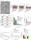
Fig. 1. Optn deficiency in oligodendrocyte and myeloid lineages promotes axonal loss and dysmyelination in the spinal cords of Optn−/− mice
(A) Top: Toluidine blue staining sections from the ventrolateral lumbar spinal cords of WT and Optn−/− mice. The brackets showing axons in the ventrolateral lumbar spinal cord white matter, and the motor neurons in the ventral lumbar spinal cord grey matter, respectively. Bottom, electron microscopic analysis of motor axonal myelination in the ventrolateral lumbar spinal cords from WT and Optn−/− mice. (B–D; F–I) The mean axonal numbers, mean g-ratios and mean axonal diameters, individual g-ratios distribution and distributions of axonal diameters in the ventrolateral lumbar spinal cord white matter (L1–L4) of WT, Optn−/− mice, OptnF/F mice, OptnF/F; Cnp-cre mice, OptnF/F; Lyz2-cre mice, OptnF/F; Gfap-cre mice and OptnF/F; Mnx1-cre as indicated. (E) The number of TUNEL+ cells in the lumbar spinal cords (L1–L4, one section each) of indicated genotype (5 mice each genotype).
Fig. 2
Fig. 2. OPTN deficiency sensitizes to necroptosis
(A) Murine primary oligodendrocytes of indicated genotypes were treated with mTNFα (10 ng/mL) +/or- Nec-1s (10 μM) for 24 hrs and cell death was assessed by using Toxilight assay (Lonza). Data are represented as the normalized means ± SEM, n=5–9 replicates per group. (B) The spinal cords of WT and Optn−/− mice were extracted with urea buffer and analyzed by western blotting using indicated abs. (C) The spinal cord lysates extracted with RIPA buffer were immunoprecipitated using anti-OPTN or anti-RIPK1 and the immunocomplexes were analyzed by western blotting using using indicated abs. (D) The spinal cords from mice of indicated genotypes were lysed in 6M urea and immunoprecipitated using anti-K48 ubiquitin chain abs. The isolated immunocomplexes and input were analyzed by western blotting with anti-RIPK1. (E) The mRNA levels of RIPK1 in the spinal cords with indicated genotypes were measured by qRT-PCR. (F) WT and Optn−/− MEFs were treated with CHX (2 μg/ml) for indicated periods of time and the lysates were analyzed by western blotting using indicated antibodies. (G) Microglia from newborns of indicated genotypes were extracted in TX114 buffer and the western blots were probed with anti-RIPK1 p-S14/15 phosphorylation and anti-RIPK1. (H) The cytokine profiles in the spinal cords were measured using a cytokine array by ELISA. (I) Heatmap of top 71 genes in the module ME1 differentially expressed in microglia of indicated genotypes. Low expression is shown in blue and high expression in red.
Fig. 3
Fig. 3. RIPK1 and RIPK3 mediate axonal pathology in the spinal cords of Optn−/− mice
(A) Dysmyelination in the spinal cords of Optn−/− mice was blocked by genetically inhibiting RIPK1 in Optn−/−; Ripk1D138N/D138N mice, pharmacologically inhibiting RIPK1 by Nec-1s (oral dosing of Nec-1s for one month starting from 8 weeks of age) and by loss of RIPK3 in Optn−/−; Ripk3−/− mice. (B–D) Mean axonal numbers, g-ratios and axonal diameters (B), individual g-ratio distributions (C), and axonal diameter distributions (D). (E) The number of TUNEL+ cells in the lumbar spinal cords (L1–L4, one section each) of indicated genotypes at 3 months of age (5 mice per genotype). (FH) Mice of indicated genotypes were tested in Open Field Test for spontaneous motor activity. The mice were at 3 months of age and 28–32 g of body weight (no statistical significant difference on body weight between different groups). The total distance traveled in one hour showed no difference between different groups (F). Optn−/− mice showed a significant deficit on the vertical rearing activity (frequency with which the mice stood on their hind legs). This deficit was blocked after dosing with Nec-1s for one month starting from 8 weeks old and in Optn−/−; Ripk1D138N/D138N double mutant mice and reduced in Optn−/−; Ripk3−/− double mutant mice (G–H).
Fig. 4
Fig. 4. RIPK1 and RIPK3 mediated axonal pathology is a common mechanism in ALS
(A) Urea buffer lysates of spinal cords from WT and SOD1G93A transgenic mice (12 weeks of age) were analyzed by western blotting using indicated abs. (BC) The myelination morphology (top), mean axonal numbers (bottom), mean g-ratios (bottom), mean axonal diameters (bottom) of the ventrolateral lumbar spinal cord white matter of SOD1G93A mice, SOD1G93A; Ripk3−/− mice (12 weeks of age) and SOD1G93A mice dosed with vehicle or Nec-1s for one month starting from 8 weeks of age. (DE) RIPK3 deficiency (D) and inhibition of RIPK1 by Nec-1s starting from 8 weeks of age (E) delayed the onset of motor dysfunction in SOD1G93A mice. (F) Sections of pathological spinal cords from human control and an ALS patient were stained with luxol fast blue for myelin to show reduced myelination in the lateral column of lower spinal cords of ALS. (G) Western blotting analysis of human control and ALS spinal cord samples using indicated abs (top) and the quantitation of RIPK1, RIPK3 and MLKL levels from 10 controls and 13 ALS cases (bottom).

Comment in

References

    1. Beeldman E, et al. A Dutch family with autosomal recessively inherited lower motor neuron predominant motor neuron disease due to optineurin mutations. Amyotroph Lateral Scler Frontotemporal Degener. 2015;16:410–411. - PubMed
    1. Cirulli ET, et al. Exome sequencing in amyotrophic lateral sclerosis identifies risk genes and pathways. Science. 2015;347:1436–1441. - PMC - PubMed
    1. Maruyama H, et al. Mutations of optineurin in amyotrophic lateral sclerosis. Nature. 2010;465:223–226. - PubMed
    1. Munitic I, et al. Optineurin insufficiency impairs IRF3 but not NF-kappaB activation in immune cells. J Immunol. 2013;191:6231–6240. - PMC - PubMed
    1. Zhu G, et al. Optineurin negatively regulates TNFalpha- induced NF-kappaB activation by competing with NEMO for ubiquitinated RIP. Curr Biol. 2007;17:1438–1443. - PubMed

Publication types

MeSH terms