. 2016 Aug 23;113(34):E4956-65.
doi: 10.1073/pnas.1525082113. Epub 2016 Aug 4.

Kinetic and thermodynamic framework for P4-P6 RNA reveals tertiary motif modularity and modulation of the folding preferred pathway

Affiliations

Kinetic and thermodynamic framework for P4-P6 RNA reveals tertiary motif modularity and modulation of the folding preferred pathway

Namita Bisaria et al. Proc Natl Acad Sci U S A. .

Abstract

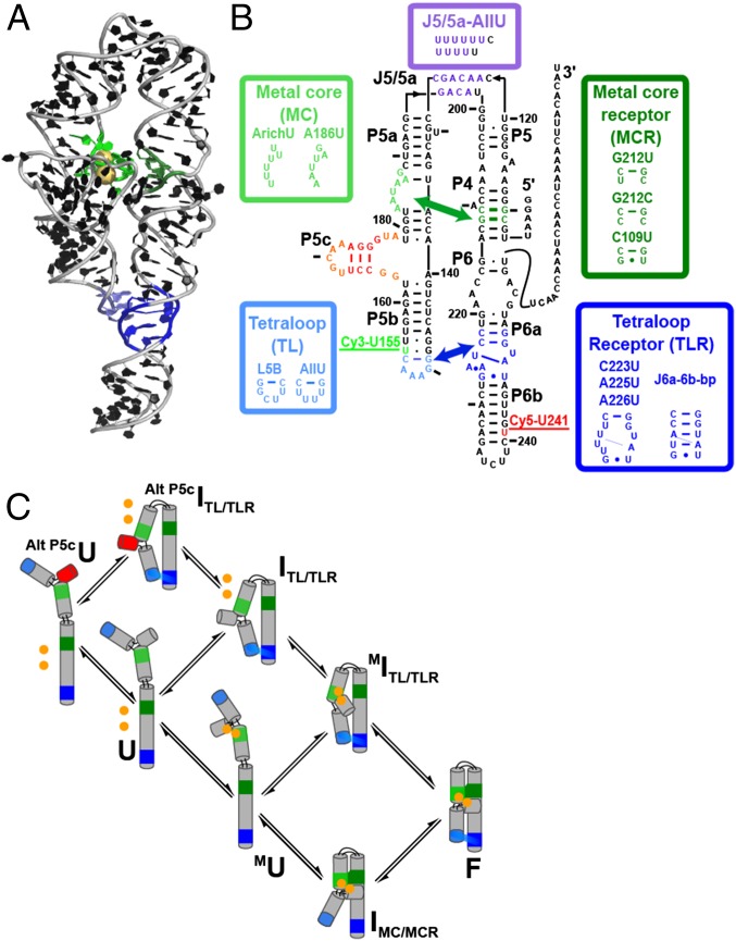
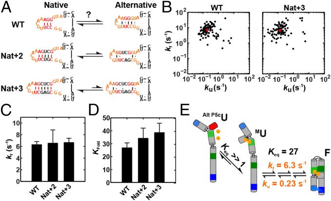
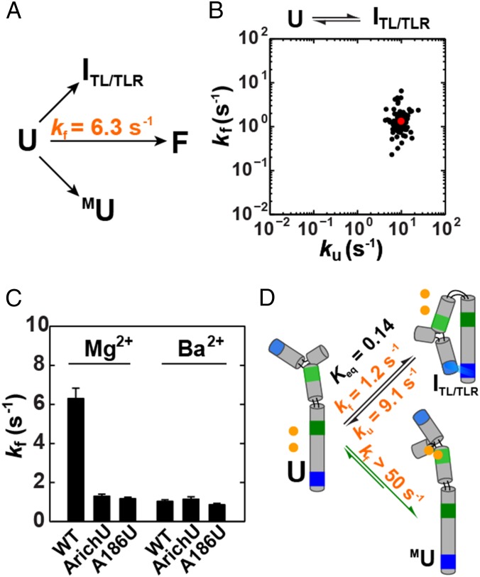
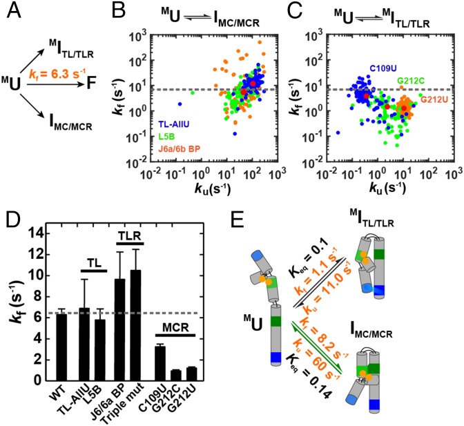
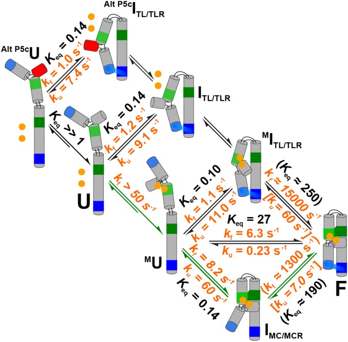
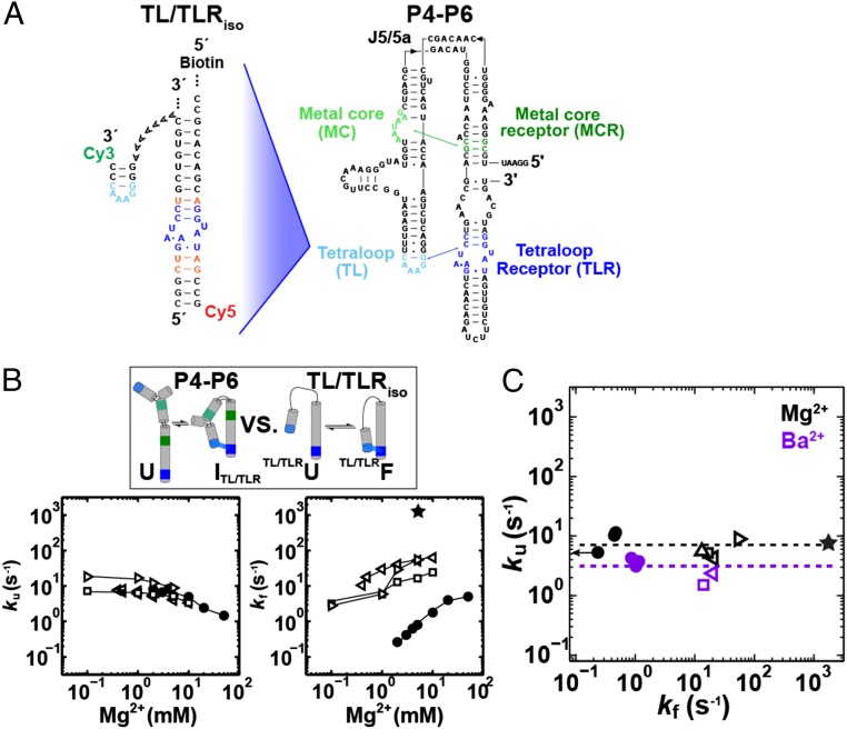
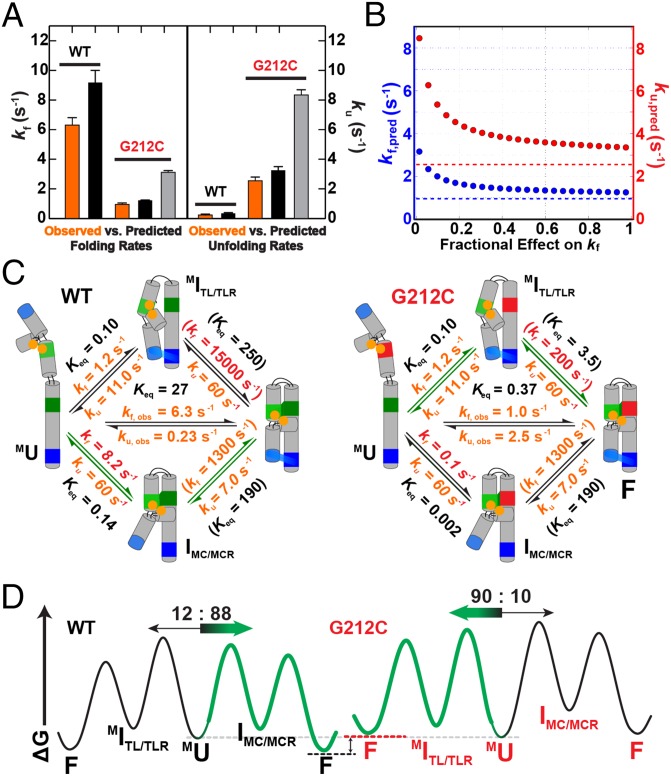
The past decade has seen a wealth of 3D structural information about complex structured RNAs and identification of functional intermediates. Nevertheless, developing a complete and predictive understanding of the folding and function of these RNAs in biology will require connection of individual rate and equilibrium constants to structural changes that occur in individual folding steps and further relating these steps to the properties and behavior of isolated, simplified systems. To accomplish these goals we used the considerable structural knowledge of the folded, unfolded, and intermediate states of P4-P6 RNA. We enumerated structural states and possible folding transitions and determined rate and equilibrium constants for the transitions between these states using single-molecule FRET with a series of mutant P4-P6 variants. Comparisons with simplified constructs containing an isolated tertiary contact suggest that a given tertiary interaction has a stereotyped rate for breaking that may help identify structural transitions within complex RNAs and simplify the prediction of folding kinetics and thermodynamics for structured RNAs from their parts. The preferred folding pathway involves initial formation of the proximal tertiary contact. However, this preference was only ∼10 fold and could be reversed by a single point mutation, indicating that a model akin to a protein-folding contact order model will not suffice to describe RNA folding. Instead, our results suggest a strong analogy with a modified RNA diffusion-collision model in which tertiary elements within preformed secondary structures collide, with the success of these collisions dependent on whether the tertiary elements are in their rare binding-competent conformations.

Keywords: RNA folding; RNA tertiary motifs; folding pathways; kinetics; single-molecule FRET.


Conflict of interest statement

The authors declare no conflict of interest.

Figures

Fig. 1.
Fig. 1.
P4-P6 RNA and proposed framework. (A) P4-P6 crystal structure (PDB ID code 1GID). Tertiary contacts are colored as follows: tetraloop (TL, light blue), tetraloop receptor (TLR, dark blue), metal core (MC, light green), metal-core receptor (MCR, dark green). Crystallographically and functionally defined Mg2+ ions bound by the MC are shown as gold spheres (46, 50, 82). (B) P4-P6 secondary structure and variants used in this study to isolate specific folding intermediates (see also Fig. 2A). Tertiary contacts are colored as in A. The positions of the donor and acceptor dyes are shown. (C) Proposed thermodynamic and kinetic framework based on previously identified structural intermediates. P4-P6 is shown in a schematic depiction, color-coded as in A and B, and tertiary contacts are shown throughout as a green swatch and a blue swatch for the MC/MCR and TL/TLR, respectively. Intermediates include the following: Alt P5cU, unfolded state with an alternative P5c secondary structure; Alt P5cITL/TLR, intermediate with the TL/TLR interaction formed and the alternative P5c secondary structure; U, unfolded state with the native P5c secondary structure (states not notated “Alt P5c” have native P5c secondary structure); ITL/TLR, intermediate with only the TL/TLR interaction formed; MU, unfolded state with the MC formed through binding of two Mg2+ ions (depicted as two bound gold spheres); MITL/TLR, intermediate in which the TL/TLR interaction and the MC are formed; IMC/MCR, intermediate with the MC/MCR interaction formed; and F, the native folded state in which both tertiary contacts are formed (49, 56, 60, 77, 82, 118).
Fig. 2.
Fig. 2.
Overall folding of WT P4-P6 and constructs with the native P5c secondary structure enforced. (A) The local equilibrium between the native and alternative P5c secondary structure of WT P4-P6 and constructs with the native P5c secondary structure enforced by two (Nat+2) or three (Nat+3) base pairs (46, 50, 77, 82). (B) Scatter plot of folding versus unfolding rate constants for WT and Nat+3 P4-P6. Data for 100 randomly selected molecules with the median folding and unfolding rate constants denoted by red circles (Dataset S1). See SI Appendix, Fig. S1 for data for a variant with the alternative P5c secondary structure (Alt+2). (C and D) Comparison of the folding rate (C) and equilibrium (D) constants of WT, Nat+2, and Nat+3. Error bars correspond to the bootstrap-estimated 95% confidence intervals (SD = 2σbootstrap). See SI Appendix, Table S1 for data and SI Appendix, Supporting Methods for experimental details. Conditions were as follows: 50 mM MOPS, pH 8.0, 5 mM MgCl2, and 100 mM KCl at 23 °C. (E) Overall P4-P6 folding occurs from an unfolded state with native P5c secondary structure (MU).
Fig. 3.
Fig. 3.
Does the folding pathway proceed via formation of the tetraloop/tetraloop receptor (TL/TLR) or the metal core (MC)? (A) Folding from U could occur either through ITL/TLR or MU. (B) Scatter plot of the folding versus unfolding rate constants of the ArichU mutant (Fig. 1C) that isolates the U-to-ITL/TLR transition. (C) Comparison of the folding rate constants of WT, ArichU, and A186U P4-P6 in Mg2+ and Ba2+. Prior work has shown that Ba2+ does not bind to the metal core so the same folding behavior is predicted for WT P4-P6 and the ArichU and A186U mutants in Ba2+ (81). This expectation was met, suggesting that there are no unintended effects from the above mutations. Errors as in Fig. 2. Conditions were as follows: 50 mM MOPS, pH 8.0, 5 mM MgCl2 or BaCl2, and 100 mM KCl at 23 °C. (D) P4-P6 folds predominantly via formation of the MU intermediate (green arrows). The equilibrium and kinetic values shown for the U-to-ITL/TLR pathway are average values from A186U and ArichU variants (SI Appendix, Table S1). Estimation of the limit for the U-to-MU transition of >50 s−1 was determined from previous literature data (SI Appendix, SI Text).
Fig. 4.
Fig. 4.
Does folding proceed from MU via formation of the tetraloop/tetraloop receptor (TL/TLR) or the metal core/metal-core receptor (MC/MCR)? (A) From MU, folding can proceed through MITL/TLR or IMC/MCR. (B) Scatter plot of the folding versus unfolding rate constants for the TL-AllU (blue), L5B (green), and J6a/6b BP (orange) mutants that destabilizes the TL/TLR; and (C) MCR variants C109U (blue), G212C (green), and G212U (orange) that destabilize the formation of the MC/MCR contact (SI Appendix, Fig. S5). (D) Comparison of the folding rate constants of WT P4-P6 with those of the TL, TLR, and MCR mutants. Triple mut, the A225U/A226U/C223U variant. The dotted gray lines in C and D indicate the rate constant for WT P4-P6 folding. The values are from SI Appendix, Table S1, and the error estimates and conditions are as in Fig. 3. (E) Preferential folding via IMC/MCR. The rate constants for the MU-to-MITL/TLR transition are for the G212U mutant (SI Appendix, Table S1) because this mutant has rate constants and behavior similar to MC mutations that ablate the MC/MCR interaction (56). The rate constants for the MU-to-IMC/MCR transition are averages of the values for the TL and TLR variants in D.
Fig. 5.
Fig. 5.
Thermodynamic and kinetic framework of P4-P6 folding (5 mM MgCl2, 100 mM KCl, 23 °C). The preferred folding pathway is depicted with the green arrows. Values for each step are taken from results in Fig. 3D, Fig. 4E, and SI Appendix, Fig. S1. The value in the square bracket for F→IMC/MCR is the average of the unfolding rate constants determined for constructs that isolate the TL/TLR (SI Appendix, Table S2). The value in the square bracket for F → MITL/TLR is the average of unfolding rate constants for constructs that isolate the MC/MCR (P4-P6, MU⇌IMC/MCR) (SI Appendix, Table S1); (MC/MCRiso, 5 mM Mg2+) (SI Appendix, Table S3). The value for Alt P5cITL/TLRAlt P5cU was corrected by a factor of two to account for the uniform twofold slower unfolding in 5 mM Ba2+ relative to 5 mM Mg2+ (Fig. 6C and SI Appendix, Table S1) because the experiment was carried out with Ba2+ to prevent Mg2+ occupancy of the metal core (MC) (81). Values in parentheses were calculated from completion of the thermodynamic cycle (SI Appendix, Supporting Methods).
Fig. 6.
Fig. 6.
Folding kinetics of tertiary contacts within different structural contexts. (A) Construct used to isolate the folding and unfolding of the TL/TLR (TL/TLRiso). Residues are colored as in Fig.1, and residues that are the same as in P4-P6 are colored orange. (B, Top) Constructs that isolate TL/TLR formation (see SI Appendix, Fig. S2 for TL/TLRiso sequences). (Bottom) Unfolding (Left) and folding (Right) rate constants for ArichU P4-P6 variant (●) and TL/TLRiso constructs (A7 linker, □; T14 linker, left triangle; and an extended tetraloop helix, right triangle) across a range of Mg2+ concentrations. Published data for TL/TLRiso are shown (A7 linker, □) (100). Lines are shown to guide the eye (published data for the TL/TLRiso were taken with 100 mM NaCl instead of 100 mM KCl; see SI Appendix, Tables S1 and S3 for values and condition details). (C) Unfolding versus folding rate constants of P4-P6 variants (closed circles: ArichU, A186U, and J5/5a AllU/Arichu) and TL/TLRiso constructs (open symbols, as in B, and including a P4-P6 helical context, △) in 5 mM Mg2+ (black) or Ba2+ (purple). The folding rate constant for J5/5a AllU/ArichU P4-P6 is an upper limit indicated by the left arrow. The folding rate constant for IMC/MCR→F (★) was calculated as described in the SI Appendix, Supporting Methods, Eq. 3. The average unfolding rates in 5 mM Mg2+ or Ba2+ are shown as black and purple dashed lines, respectively, and were twofold slower in Ba2+ than Mg2+. Data are from SI Appendix, Tables S1–S3.
Fig. 7.
Fig. 7.
The kinetic effects of the G212C MCR mutation. (A) Comparison of the observed (orange) versus predicted (black and gray) overall folding (Left) and unfolding (Right) rate constants of WT P4-P6 and G212C P4-P6 (SI Appendix, Supporting Methods). For WT, the predicted rate constants are calculated from the values in Fig. 5 that are reproduced in C of this figure. For G212C, the predicted folding and unfolding rate constants assume the full equilibrium effect of the mutation either on the folding (black) or unfolding (gray) rate constant for the MC/MCR interaction. (B) Predicted values for the overall folding (blue) and unfolding (red) rate constants for the G212C mutant over a range of hypothetical MC/MCR folding and unfolding rate constants, assuming that the full equilibrium effect of the mutation (72-fold) is apportioned between these rate constants, ranging from the full effect of the mutation on the unfolding rate constant [(fractional effect on kf) = 0] to the full effect on the folding rate constant [(fractional effect on kf) = 1]. The observed overall folding and unfolding rate constants for G212C are represented by the dashed lines. (C) Simplified folding framework for WT P4-P6 (Left; values from Fig. 5) and G212C P4-P6 (Right). Values for G212C P4-P6 are the same as for WT except for steps for MC/MCR formation, which are decreased by the full equilibrium effect of the mutation (72-fold). (D) Free energy diagrams for WT P4-P6 (Left) and the G212C mutant (Right) folding. The preferred folding pathway is shown in green, and the flux through each pathway is given above each pathway and is designated by the relative arrow size. Profiles are to scale.

References

    1. Warf MB, Berglund JA. Role of RNA structure in regulating pre-mRNA splicing. Trends Biochem Sci. 2010;35(3):169–178. - PMC - PubMed
    1. Rinn JL, Chang HY. Genome regulation by long noncoding RNAs. Annu Rev Biochem. 2012;81(1):145–166. - PMC - PubMed
    1. Mercer TR, Mattick JS. Structure and function of long noncoding RNAs in epigenetic regulation. Nat Struct Mol Biol. 2013;20(3):300–307. - PubMed
    1. Cech TR, Steitz JA. The noncoding RNA revolution-trashing old rules to forge new ones. Cell. 2014;157(1):77–94. - PubMed
    1. Staley JP, Guthrie C. Mechanical devices of the spliceosome: Motors, clocks, springs, and things. Cell. 1998;92(3):315–326. - PubMed

Publication types

LinkOut - more resources