Review
. 2016 Nov;100(5):969-978.
doi: 10.1189/jlb.4MR0216-079R. Epub 2016 Aug 4.

Autophagy in leukocytes and other cells: mechanisms, subsystem organization, selectivity, and links to innate immunity

Affiliations
Review

Autophagy in leukocytes and other cells: mechanisms, subsystem organization, selectivity, and links to innate immunity

Vojo Deretic. J Leukoc Biol. 2016 Nov.

Abstract

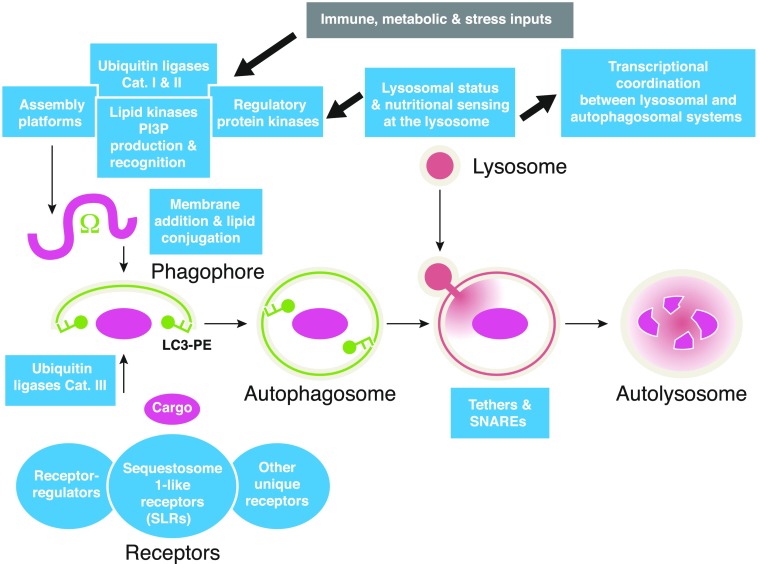
Autophagy is a fundamental biologic process that fulfills general and specialized roles in cytoplasmic homeostasis. The cell-autonomous antimicrobial functions of autophagy have been established in the macrophage. These cells and other leukocytes continue to be the cells of choice in studying autophagy in immunity and inflammation. This review uses several model examples that will be of interest to leukocyte and cell biologists alike. Furthermore, it comprehensively covers the subsystems in autophagy as they apply to all mammalian cells and incorporates the recent progress in our understanding of how these modules come together-a topic that should be of interest to all readers.

Keywords: ATG; Crohn's disease; IRGM; TRIM; tuberculosis.


Figures

Figure 1.
Figure 1.. Autophagy pathway with its regulatory and execution subsystems.
Depicted are the simplified conventional views of canonical macroautophagy organelles and pathway with different subsystems detailed in Table 1. Ω, Omegasome, a PI3P-positive structure on the ER believed to provide a cradle for formation of phagophores positive for mAtg8 orthologs (e.g., LC3-PE, also known as LC3-II). Phagophores enwrap the cargo, enlarge, and close to form double-membrane autophagosomes that then fuse with lysosomes to form autolysosomes, where the captured cargo is degraded. Cargo is brought into the phagophores via receptors that recognize cargo through binding to tags, such as ubiquitin, placed on the cargo by E3 ubiquitin ligases (category III; see Table 1). Protein kinases, lipid kinases, and assembly platforms (detailed in Table 1) drive the formation of autophagosomes. These are regulated by E3 ubiquitin ligases that either stabilize (category I) or destabilize (category II) them. The entire system is regulated by signals that can be immunologic, nutritional, differentiation in nature, or from various stressors. Lysosome is not just a passive contributor of lysosomal contents but also works as a sensory organelle that relays the status to the nucleus, whereby autophagosomal and lysosomal biogenesis are coordinated at the transcriptional level.

References

    1. Mizushima N., Komatsu M. (2011) Autophagy: renovation of cells and tissues. Cell 147, 728–741. - PubMed
    1. Ashford T. P., Porter K. R. (1962) Cytoplasmic components in hepatic cell lysosomes. J. Cell Biol. 12, 198–202. - PMC - PubMed
    1. Deter R. L., De Duve C. (1967) Influence of glucagon, an inducer of cellular autophagy, on some physical properties of rat liver lysosomes. J. Cell Biol. 33, 437–449. - PMC - PubMed
    1. Deter R. L., Baudhuin P., De Duve C. (1967) Participation of lysosomes in cellular autophagy induced in rat liver by glucagon. J. Cell Biol. 35, C11–C16. - PMC - PubMed
    1. Deretic V., Kimura T., Timmins G., Moseley P., Chauhan S., Mandell M. (2015) Immunologic manifestations of autophagy. J. Clin. Invest. 125, 75–84. - PMC - PubMed

Publication types

MeSH terms