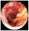
Sialoendoscopy as a treatment for an obstructed mandibular salivary duct in a horse
- PMID: 27493288
- PMCID: PMC4944566
Sialoendoscopy as a treatment for an obstructed mandibular salivary duct in a horse
Abstract
A 14-year-old Quarter Horse was examined for a draining tract of 8 months' duration on the right mandible that was non-responsive to antibiotic therapy and surgical therapy. Further investigation and subsequent treatment with sialoendoscopy and ultrasonography were performed to relieve an obstruction of plant awns in the mandibular salivary duct.
Sialo-endoscopie comme traitement pour un canal salivaire mandibulaire bloqué chez un cheval. Un cheval Quarter Horse âgé de 14 ans a été examiné pour une fistule purulente d’une durée de 8 mois à la mandibule droite qui ne répondait pas à la thérapie antibiotique et à la thérapie chirurgicale. De nouvelles investigations et le traitement subséquent à l’aide de la sialo-endoscopie et de l’échographie ont été réalisés pour éliminer un blocage du canal salivaire mandibulaire par des barbes de plantes.(Traduit par Isabelle Vallières).
Figures


References
-
- Kilcoyne I, Watson JL, Spier SJ, Whitcomb MB, Vaughan B. Septic sialoadenitis in equids: A retrospective study of 18 cases (1998–2010) Equine Vet J. 2015;47:54–59. - PubMed
-
- Baratt RM, Rawlinson JE. Diagnostic imaging in veterinary dental practice. J Am Vet Med Assoc. 2013;243:203–205. - PubMed
-
- Vaughan B, Whitcomb MB, Biscoe B. Review of ultrasonographic techniques to evaluate the equine skull and head structures. Proceedings of the 57th Annual Convention of the American Association of Equine Practitioners; San Antonio, Texas, USA. 18–22 November, 2011.
-
- Ziegler CM, Steveling H, Seubert M, Mühling J. Endoscopy: A minimally invasive procedure for diagnosis and treatment of diseases of the salivary glands: Six years of practical experience. Br J Oral Maxillofac Surg. 2004;42:1–7. - PubMed
-
- Pace CG, Hwang KG, Papadaki M, Troulis MJ. Interventional sialoendoscopy for treatment of obstructive sialadenitis. J Oral Maxillofac Surg. 2014;72:2157–2166. - PubMed
