Defense-Related Responses in Fruit of the Nonhost Chili Pepper against Xanthomonas axonopodis pv. glycines Infection
- PMID: 27493606
- PMCID: PMC4968641
- DOI: 10.5423/PPJ.OA.12.2015.0256
Defense-Related Responses in Fruit of the Nonhost Chili Pepper against Xanthomonas axonopodis pv. glycines Infection
Abstract
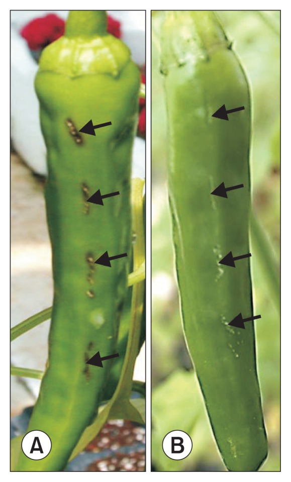
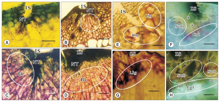
Xanthomonas axonopodis pv. glycines (Xag ) is a necrotrophic bacterial pathogen of the soybean that causes bacterial pustules and is a nonhost pathogen of the chili pepper. In the current study, chili pepper fruit wound inoculated in planta with Xag 8ra formed necrotic lesions on the fruit surface and induced several structural and chemical barriers systemically in the fruit tissue. The initial defense response included programmed cell death of necrotizing and necrotized cells, which was characterized by nuclear DNA cleavage, as detected by TUNEL-confocal laser scanning microscopy (CLSM), and phosphatidylserine exposure on cell walls distal to the infection site, as detected by Annexin V FLUOS-CLSM. These two responses may facilitate cell killing and enhance transportation of cell wall materials used for cell wall thickening, respectively. The cells beneath the necrotic tissue were enlarged and divided to form periclinal cell walls, resulting in extensive formation of several parallel boundary layers at the later stages of infection, accompanying the deposition of wall fortification materials for strengthening structural defenses. These results suggest that nonhost resistance of chili pepper fruit against the nonhost necrotrophic pathogen Xag 8ra is activated systematically from the initial infection until termination of the infection cycle, resulting in complete inhibition of bacterial pathogenesis by utilizing organ-specific in situ physiological events governed by the expression of genes in the plant fruit organ.
Keywords: Xanthomonas axonopodis pv. glycines; chili pepper; hypersensitive response; programmed cell death.
Figures


Similar articles
-
Insight into Types I and II nonhost resistance using expression patterns of defense-related genes in tobacco.Planta. 2006 Apr;223(5):1101-7. doi: 10.1007/s00425-006-0232-1. Epub 2006 Feb 16. Planta. 2006. PMID: 16482435
-
EST and microarray analyses of pathogen-responsive genes in hot pepper (Capsicum annuum L.) non-host resistance against soybean pustule pathogen (Xanthomonas axonopodis pv. glycines).Funct Integr Genomics. 2004 Jul;4(3):196-205. doi: 10.1007/s10142-003-0099-1. Epub 2004 Feb 4. Funct Integr Genomics. 2004. PMID: 14760538
-
[Xanthomonas axonopodis pv. glycines Zur is involved in pathogenicity in host and hypersensitive responses in nonhosts].Sheng Wu Gong Cheng Xue Bao. 2024 Oct 25;40(10):3603-3618. doi: 10.13345/j.cjb.230816. Sheng Wu Gong Cheng Xue Bao. 2024. PMID: 39467753 Chinese.
-
Final report on the safety assessment of capsicum annuum extract, capsicum annuum fruit extract, capsicum annuum resin, capsicum annuum fruit powder, capsicum frutescens fruit, capsicum frutescens fruit extract, capsicum frutescens resin, and capsaicin.Int J Toxicol. 2007;26 Suppl 1:3-106. doi: 10.1080/10915810601163939. Int J Toxicol. 2007. PMID: 17365137 Review.
-
Nonhost resistance against bacterial pathogens: retrospectives and prospects.Annu Rev Phytopathol. 2013;51:407-27. doi: 10.1146/annurev-phyto-082712-102319. Annu Rev Phytopathol. 2013. PMID: 23725473 Review.
Cited by
-
An improved bacterial mRNA enrichment strategy in dual RNA sequencing to unveil the dynamics of plant-bacterial interactions.Plant Methods. 2024 Jul 1;20(1):99. doi: 10.1186/s13007-024-01227-x. Plant Methods. 2024. PMID: 38951818 Free PMC article.
References
-
- Agrios GN. Plant pathology. 5th ed. Academic Press; San Diego, CA, USA: 2005.
-
- Biggs AR. Phellogen regeneration in injured peach tree bark. Ann Bot. 1986;57:463–470.
-
- Biggs AR. Temporal changes in the infection court after wounding of peach bark are associated with cultivar variation in infection by Leucostoma persoonii. Phytopathology. 1989;79:627–630. doi: 10.1094/Phyto-79-627. - DOI
-
- Chen LQ, Hou BH, Lalonde S, Takanaga H, Hartung ML, Qu XQ, Guo WJ, Kim JG, Underwood W, Chaudhuri B, Chermak D, Antony G, White FF, Somerville SC, Mudgett MB, Frommer WB. Sugar transporters for intercellular exchange and nutrition of pathogens. Nature. 2010;468:527–532. doi: 10.1038/nature09606. - DOI - PMC - PubMed
LinkOut - more resources
Full Text Sources
Other Literature Sources