Review
. 2016 Jul 21:7:306.
doi: 10.3389/fphys.2016.00306. eCollection 2016.

"Decoding" Angiogenesis: New Facets Controlling Endothelial Cell Behavior

Affiliations
Review

"Decoding" Angiogenesis: New Facets Controlling Endothelial Cell Behavior

Raj Sewduth et al. Front Physiol. .

Abstract

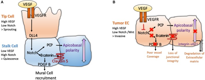
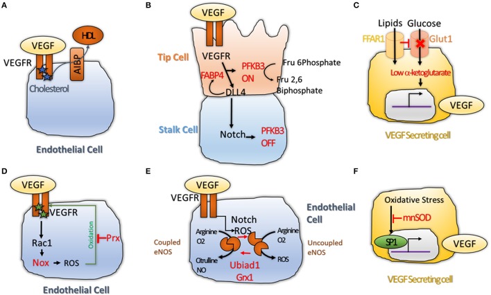
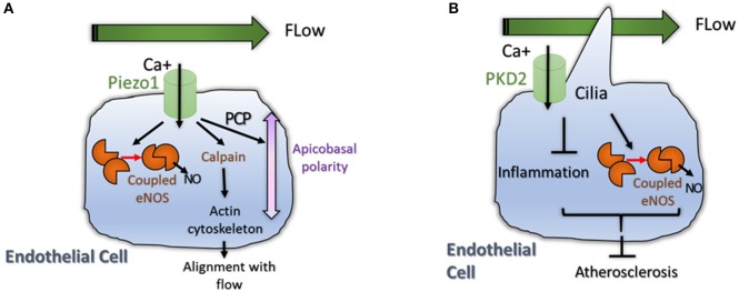
Angiogenesis, the formation of new blood vessels, is a unique and crucial biological process occurring during both development and adulthood. A better understanding of the mechanisms that regulates such process is mandatory to intervene in pathophysiological conditions. Here we highlight some recent argument on new players that are critical in endothelial cells, by summarizing novel discoveries that regulate notorious vascular pathways such as Vascular Endothelial Growth Factor (VEGF), Notch and Planar Cell Polarity (PCP), and by discussing more recent findings that put metabolism, redox signaling and hemodynamic forces as novel unforeseen facets in angiogenesis. These new aspects, that critically regulate angiogenesis and vascular homeostasis in health and diseased, represent unforeseen new ground to develop anti-angiogenic therapies.

Keywords: Notch signaling pathway; angiogenesis; flow dynamics; metabolism; redox signaling.


Figures

Figure 1
Figure 1
(A) Interplay between VEGF and Notch signaling is important for tip and stalk cell differentiation in physiological angiogenesis. The tip cell is characterized by high VEGF and low Notch levels, inducing sprouting. The stalk cell expresses low VEGF and high Notch, leading to activation of B catenin that stabilizes junctions and enables mural cell recruitment (Gavard and Gutkind, ; Reis et al., 2012). High Notch signaling also enables polarization of the stalk cell, a process important for lumenization (Phng et al., 2009); (B) The balance between VEGF and Notch signaling is lost in tumor endothelium. High VEGF and low Notch makes the tumor endothelium invasive and unstable. Loss of Notch downregulates Wnt canonical and non-canonical pathway, disrupting junctional integrity and vessel coverage (Fan et al., ; Chatterjee et al., 2013). High VEGF promotes sprouting and formation of podosomes at the basal side of the cell, a structure important for degradation of the extracellular matrix and cell motility (Seano et al., 2014).
Figure 2
Figure 2
(A) Cholesterol maintains VEGFR2 active as dimers. Transfer of Cholesterol to HDL through the AIBP lipid transporter makes VEGFR2 inactive by inducing its monomerization (Avraham-Davidi et al., 2012); (B) VEGF and Notch interplay regulates Phosphofructokinase-2/Fructose-2,6-Bisphosphatase 3 (PFKFB3) activity. PFKB3 is active in tip cell, where glycolysis is promoted to favor sprouting of the tip cell. While in the stalk cell, High Notch levels shuts down PFKB3 activity, leading to quiescence (De Bock et al., ; Schoors et al., 2014); (C) Free Fatty Acid Receptor 1 (FFAR1) activates VEGF-A expression. FFAR1 is a lipid transporter that down-regulates Glut1 glucose transporter expression when free lipids are available. This reduces the levels of alpha-ketoglutarate in the cell leading to activation of VEGF-A transcription (Joyal et al., 2016); (D) VEGFR2 promotes ROS production via Rac1 and Nox2 (Diebold et al., 2009). High level of ROS induce oxidation of VEGF2 on two cysteine residues, making it inactive. Antioxidant enzyme Peroxiredoxin2 (Prx2) can buffer the ROS levels to keep VEGFR2 cysteines in a reduced state to protect its activity (Kang et al., 2011); (E) Redox balance regulates the activity of endothelial Nitric Oxide Synthase (eNOS)that can produce Nitric Oxide (NO) when in coupled conformation and generates ROS (•2) when in an uncoupled conformation. Notch and ROS levels are regulators of this balance. Two antioxidant enzymes, the prenyltransferase Ubiad1 and Glutaredoxin1 (Grx1) were shown to promote shift of eNOS from uncoupled, to coupled conformation; showing that antioxidants are essential for maintenance of the NO balance and normal cell physiology (Chen et al., ; Mugoni et al., 2013); (F) ROS were shown to increase VEGF-A transcription via the transcription factor SP1 (Gonzalez-Pacheco et al., 2006); while antioxidant enzyme Manganese-dependent Superoxide Dismutase (MnSOD) was shown to block this process (Wang et al., 2005), demonstrating that redox balance regulates VEGF secretion directly.
Figure 3
Figure 3
(A) Physiological flow induces eNOS signaling and induces reorganization and polarization of the EC. A mechanosensor and calcium transporter called Piezo1 was shown to be activated by flow. High intracellular Ca+ levels induce coupling of eNOS but also activate Calpain, an enzyme important for actin cytoskeleton reorganization (Li et al., 2014). High Calcium levels also activate non canonical Wnt pathway (PCP) promoting polarization of the cell; (B) Primary Cilia is important for flow sensing in ECs. The cilium is essential for accumulation of intracellular Calcium that promote NO production by eNOS and reduces inflammation (Goetz et al., 2014). Both processes reduce atherosclerosis in aortic endothelium (Dinsmore and Reiter, 2016).

References

    1. Adams R. H., Alitalo K. (2007). Molecular regulation of angiogenesis and lymphangiogenesis. Nat. Rev. Mol. Cell Biol. 8, 464–478. 10.1038/nrm2183 - DOI - PubMed
    1. Alcayaga-Miranda F., Varas-Godoy M., Khoury M. (2016). Harnessing the angiogenic potential of stem cell-derived exosomes for vascular regeneration. Stem Cells Int. 2016:3409169. 10.1155/2016/3409169 - DOI - PMC - PubMed
    1. Ando J., Yamamoto K. (2013). Flow detection and calcium signalling in vascular endothelial cells. Cardiovasc. Res. 99, 260–268. 10.1093/cvr/cvt084 - DOI - PubMed
    1. Angius F., Uda S., Piras E., Spolitu S., Ingianni A., Batetta B., et al. . (2015). Neutral lipid alterations in human herpesvirus 8-infected HUVEC cells and their possible involvement in neo-angiogenesis. BMC Microbiol. 15:74. 10.1186/s12866-015-0415-7 - DOI - PMC - PubMed
    1. Avraham-Davidi I., Ely Y., Pham V. N., Castranova D., Grunspan M., et al. . (2012). ApoB-containing lipoproteins regulate angiogenesis by modulating expression of VEGF receptor 1. Nat. Med. 18, 967–973. 10.1038/nm.2759 - DOI - PMC - PubMed