

Differentiation of Human Adipose Derived Stem Cells into Smooth Muscle Cells Is Modulated by CaMKIIγ
- PMID: 27493668
- PMCID: PMC4963582
- DOI: 10.1155/2016/1267480
Differentiation of Human Adipose Derived Stem Cells into Smooth Muscle Cells Is Modulated by CaMKIIγ
Abstract
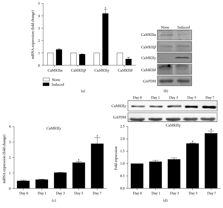
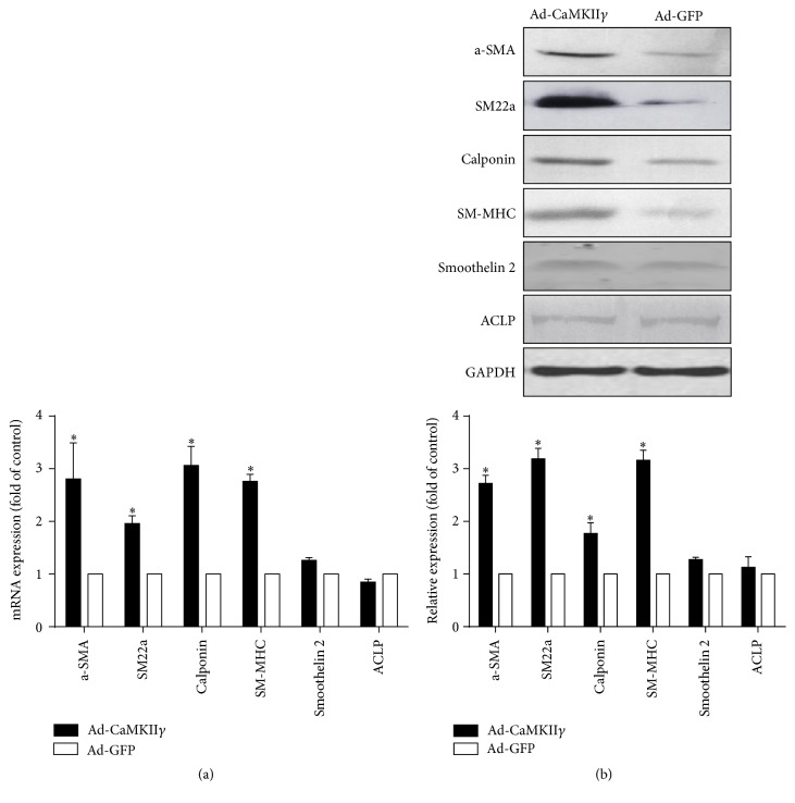
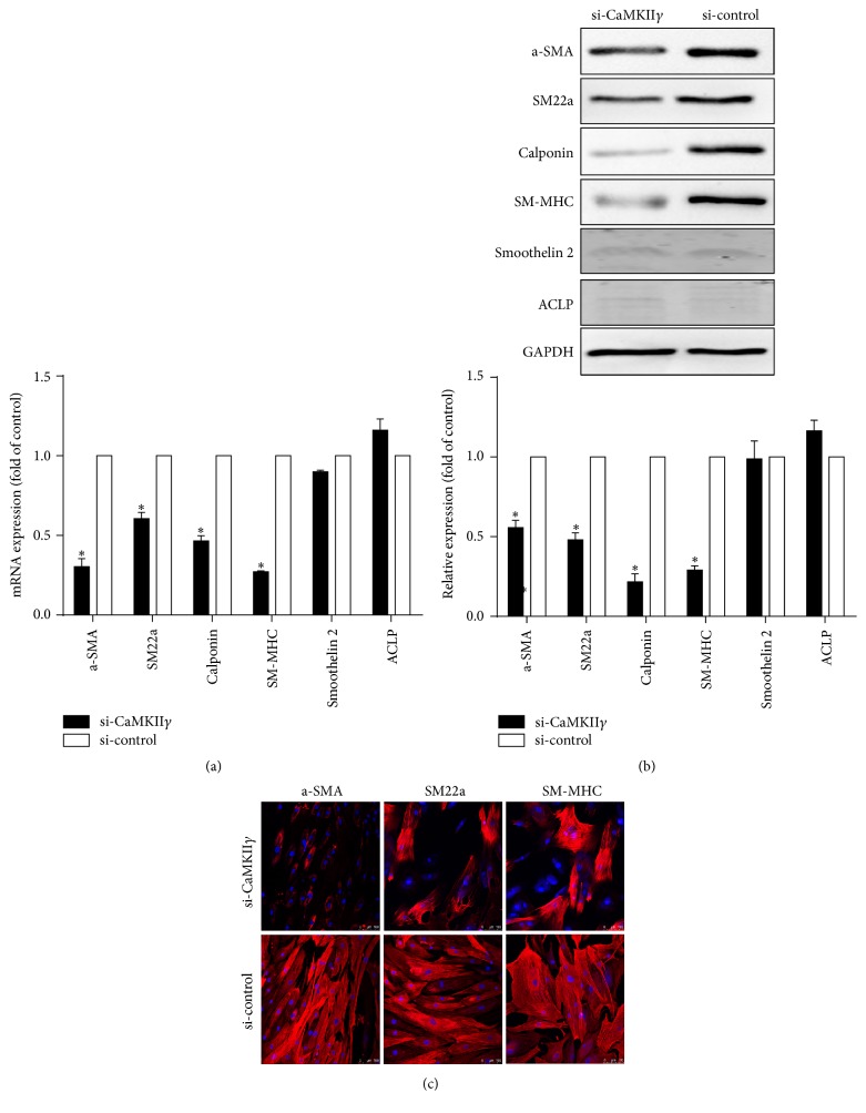
The multifunctional Ca(2+)/calmodulin-dependent protein kinase II (CaMKII) is known to participate in maintenance and switches of smooth muscle cell (SMC) phenotypes. However, which isoform of CaMKII is involved in differentiation of adult mesenchymal stem cells into contractile SMCs remains unclear. In the present study, we detected γ isoform of CaMKII in differentiation of human adipose derived stem cells (hASCs) into SMCs that resulted from treatment with TGF-β1 and BMP4 in combination for 7 days. The results showed that CaMKIIγ increased gradually during differentiation of hASCs as determined by real-time PCR and western blot analysis. The siRNA-mediated knockdown of CaMKIIγ decreased the protein levels and transcriptional levels of smooth muscle contractile markers (a-SMA, SM22a, calponin, and SM-MHC), while CaMKIIγ overexpression increases the transcriptional and protein levels of smooth muscle contractile markers. These results suggested that γ isoform of CaMKII plays a significant role in smooth muscle differentiation of hASCs.
Figures


Similar articles
-
Differentiation of adipose-derived stem cells into contractile smooth muscle cells induced by transforming growth factor-beta1 and bone morphogenetic protein-4.Tissue Eng Part A. 2010 Apr;16(4):1201-13. doi: 10.1089/ten.TEA.2009.0303. Tissue Eng Part A. 2010. PMID: 19895205
-
A small diameter elastic blood vessel wall prepared under pulsatile conditions from polyglycolic acid mesh and smooth muscle cells differentiated from adipose-derived stem cells.Biomaterials. 2010 Feb;31(4):621-30. doi: 10.1016/j.biomaterials.2009.09.086. Epub 2009 Oct 12. Biomaterials. 2010. PMID: 19819545
-
Targeting of a novel Ca+2/calmodulin-dependent protein kinase II is essential for extracellular signal-regulated kinase-mediated signaling in differentiated smooth muscle cells.Circ Res. 2005 Sep 16;97(6):541-9. doi: 10.1161/01.RES.0000182630.29093.0d. Epub 2005 Aug 18. Circ Res. 2005. PMID: 16109919
-
TGF-β1-induced differentiation of SHED into functional smooth muscle cells.Stem Cell Res Ther. 2017 Jan 23;8(1):10. doi: 10.1186/s13287-016-0459-0. Stem Cell Res Ther. 2017. PMID: 28114966 Free PMC article.
-
Ca2+/calmodulin-dependent protein kinase II-γ (CaMKIIγ) negatively regulates vascular smooth muscle cell proliferation and vascular remodeling.FASEB J. 2016 Mar;30(3):1051-64. doi: 10.1096/fj.15-279158. Epub 2015 Nov 13. FASEB J. 2016. PMID: 26567004 Free PMC article.
Cited by
-
The Role of Adipose-Derived Stem Cells, Dermal Regenerative Templates, and Platelet-Rich Plasma in Tissue Engineering-Based Treatments of Chronic Skin Wounds.Stem Cells Int. 2020 Jan 9;2020:7056261. doi: 10.1155/2020/7056261. eCollection 2020. Stem Cells Int. 2020. PMID: 32399048 Free PMC article. Review.
-
Controlled Growth Factor Delivery and Cyclic Stretch Induces a Smooth Muscle Cell-like Phenotype in Adipose-Derived Stem Cells.Cells. 2021 Nov 11;10(11):3123. doi: 10.3390/cells10113123. Cells. 2021. PMID: 34831345 Free PMC article.
-
The role of BMP4 in adipose-derived stem cell differentiation: A minireview.Front Cell Dev Biol. 2022 Oct 21;10:1045103. doi: 10.3389/fcell.2022.1045103. eCollection 2022. Front Cell Dev Biol. 2022. PMID: 36340030 Free PMC article. Review.
-
Directional Topography Influences Adipose Mesenchymal Stromal Cell Plasticity: Prospects for Tissue Engineering and Fibrosis.Stem Cells Int. 2019 May 5;2019:5387850. doi: 10.1155/2019/5387850. eCollection 2019. Stem Cells Int. 2019. PMID: 31191675 Free PMC article.
-
The deregulation of STIM1 and store operative calcium entry impaired aortic smooth muscle cells contractility in aortic medial degeneration.Biosci Rep. 2019 Jan 3;39(1):BSR20181504. doi: 10.1042/BSR20181504. Print 2019 Jan 31. Biosci Rep. 2019. PMID: 30504131 Free PMC article.
References
-
- Deslex S., Negrel R., Vannier E. J., Etienne J., Ailhaud G. Differentiation of human adipocyte precursors in a chemically defined serum-free medium. Experimental Cell Research. 2007;313:2875–2886. - PubMed
LinkOut - more resources
Full Text Sources
Other Literature Sources
Research Materials
Miscellaneous
