

Homology modeling and virtual screening studies of FGF-7 protein-a structure-based approach to design new molecules against tumor angiogenesis
- PMID: 27493695
- PMCID: PMC4958063
- DOI: 10.1007/s12154-016-0152-x
Homology modeling and virtual screening studies of FGF-7 protein-a structure-based approach to design new molecules against tumor angiogenesis
Abstract
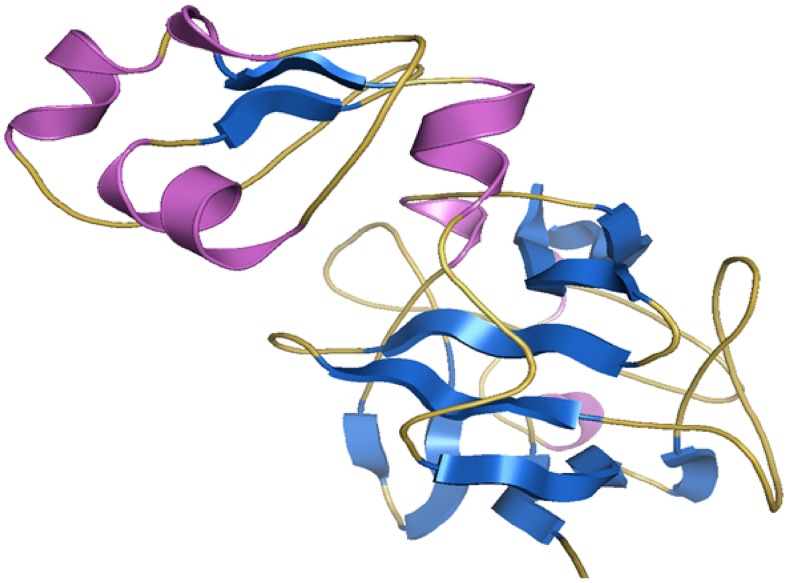
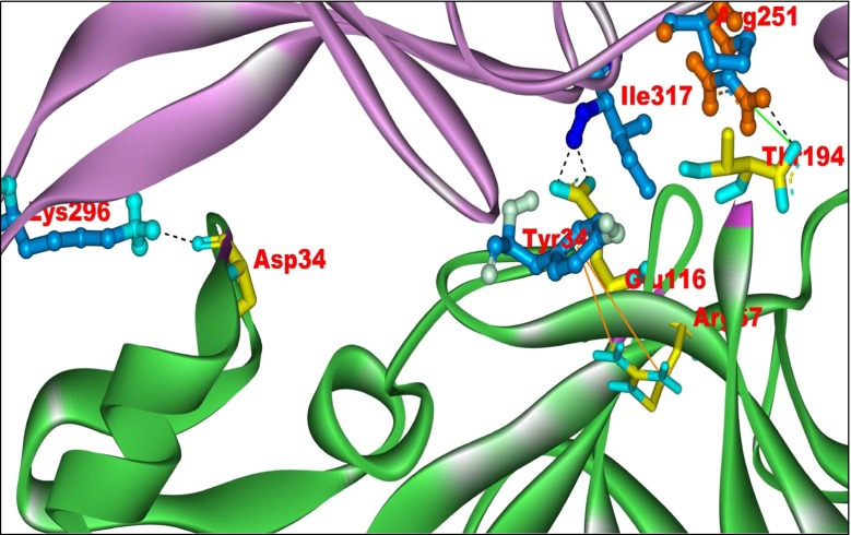
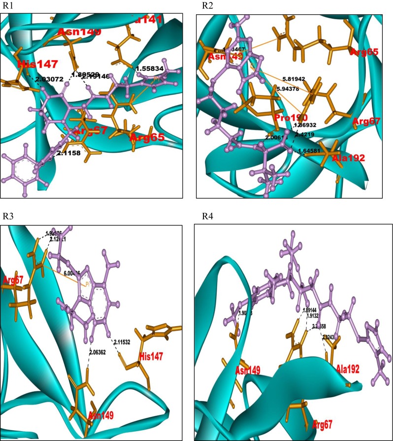
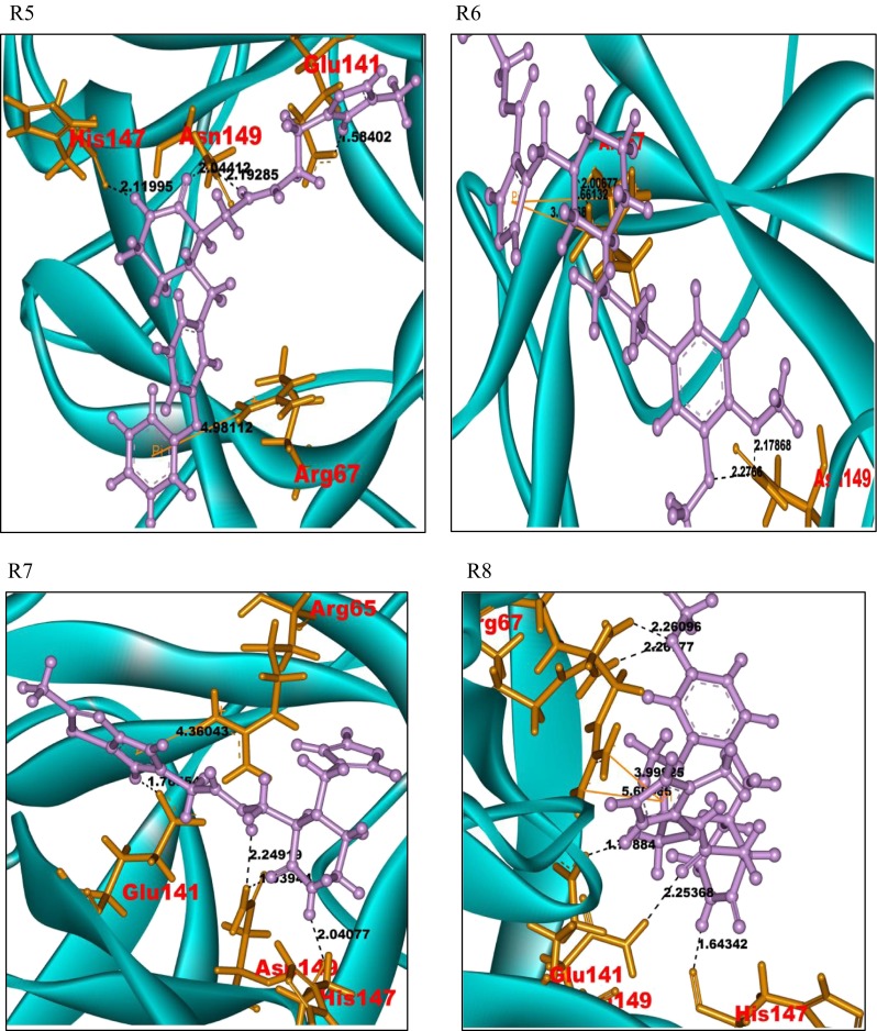
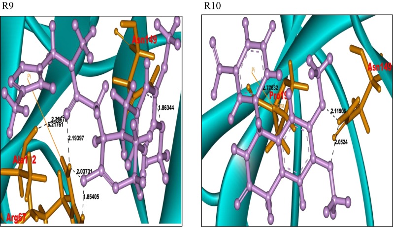
Keratinocyte growth factor (KGF) protein is a member of the fibroblast growth factor (FGF) family, which is also known as FGF-7. The FGF-7 plays an important role in tumor angiogenesis. In the present work, FGF-7 is treated as a potential therapeutic target to prevent angiogenesis in cancerous tissue. Computational techniques are applied to evaluate and validate the 3D structure of FGF-7 protein. The active site region of the FGF-7 protein is identified based on hydrophobicity calculations using CASTp and Q-site Finder active site prediction tools. The protein-protein docking study of FGF-7 with its natural receptor FGFR2b is carried out to confirm the active site region in FGF-7. The amino acid residues Asp34, Arg67, Glu116, and Thr194 in FGF-7 interact with the receptor protein (FGFR2b). A grid is generated at the active site region of FGF-7 using Glide module of Schrödinger suite. Subsequently, a virtual screening study is carried out at the active site using small molecular structural databases to identify the ligand molecules. The binding interactions of the ligand molecules, with piperazine moiety as a pharmacophore, are observed at Arg67 and Glu149 residues of the FGF-7 protein. The identified ligand molecules against the FGF-7 protein show permissible pharmacokinetic properties (ADME). The ligand molecules with good docking scores and satisfactory pharmacokinetic properties are prioritized and identified as novel ligands for the FGF-7 protein in cancer therapy.
Keywords: ADME; Angiogenesis; FGF-7; Protein–protein docking; Virtual screening.
Figures
Similar articles
-
Targeting the ubiquitin-conjugating enzyme E2D4 for cancer drug discovery-a structure-based approach.J Chem Biol. 2016 Dec 24;10(2):51-67. doi: 10.1007/s12154-016-0164-6. eCollection 2017 Apr. J Chem Biol. 2016. PMID: 28405240 Free PMC article.
-
Identification of Novel Antagonists for Rab38 Protein by Homology Modeling and Virtual Screening.Comb Chem High Throughput Screen. 2016;19(10):875-892. doi: 10.2174/1386207319666161026153237. Comb Chem High Throughput Screen. 2016. PMID: 27784220
-
Design of novel lead molecules against RhoG protein as cancer target - a computational study.J Biomol Struct Dyn. 2017 Nov;35(14):3119-3139. doi: 10.1080/07391102.2016.1244492. Epub 2016 Nov 4. J Biomol Struct Dyn. 2017. PMID: 27691842
-
Structure Elucidation and Identification of Novel Lead Molecules against Sulfur Import Protein cysA of Mycobacterium tuberculosis.Curr Protein Pept Sci. 2023;24(7):589-609. doi: 10.2174/1389203724666230713124339. Curr Protein Pept Sci. 2023. PMID: 37448368
-
Free resources to assist structure-based virtual ligand screening experiments.Curr Protein Pept Sci. 2007 Aug;8(4):381-411. doi: 10.2174/138920307781369391. Curr Protein Pept Sci. 2007. PMID: 17696871 Review.
Cited by
-
Construction of transplantable artificial vascular tissue based on adipose tissue-derived mesenchymal stromal cells by a cell coating and cryopreservation technique.Sci Rep. 2021 Sep 9;11(1):17989. doi: 10.1038/s41598-021-97547-2. Sci Rep. 2021. PMID: 34504254 Free PMC article.
-
Targeting the ubiquitin-conjugating enzyme E2D4 for cancer drug discovery-a structure-based approach.J Chem Biol. 2016 Dec 24;10(2):51-67. doi: 10.1007/s12154-016-0164-6. eCollection 2017 Apr. J Chem Biol. 2016. PMID: 28405240 Free PMC article.
-
Induced clustering of SHP2-depleted tumor cells in vascular islands restores sensitivity to MEK/ERK inhibition.J Clin Invest. 2025 Mar 25;135(10):e181609. doi: 10.1172/JCI181609. eCollection 2025 May 15. J Clin Invest. 2025. PMID: 40131370 Free PMC article.
-
Micro-RNA193a-3p Inhibits Breast Cancer Cell Driven Growth of Vascular Endothelial Cells by Altering Secretome and Inhibiting Mitogenesis: Transcriptomic and Functional Evidence.Cells. 2022 Sep 23;11(19):2967. doi: 10.3390/cells11192967. Cells. 2022. PMID: 36230929 Free PMC article.
References
LinkOut - more resources
Full Text Sources
Other Literature Sources
