Review
. 2016 Jul 12:7:2041731416650838.
doi: 10.1177/2041731416650838. eCollection 2016 Jan-Dec.

Methods for culturing retinal pigment epithelial cells: a review of current protocols and future recommendations

Affiliations
Review

Methods for culturing retinal pigment epithelial cells: a review of current protocols and future recommendations

Aaron H Fronk et al. J Tissue Eng. .

Abstract

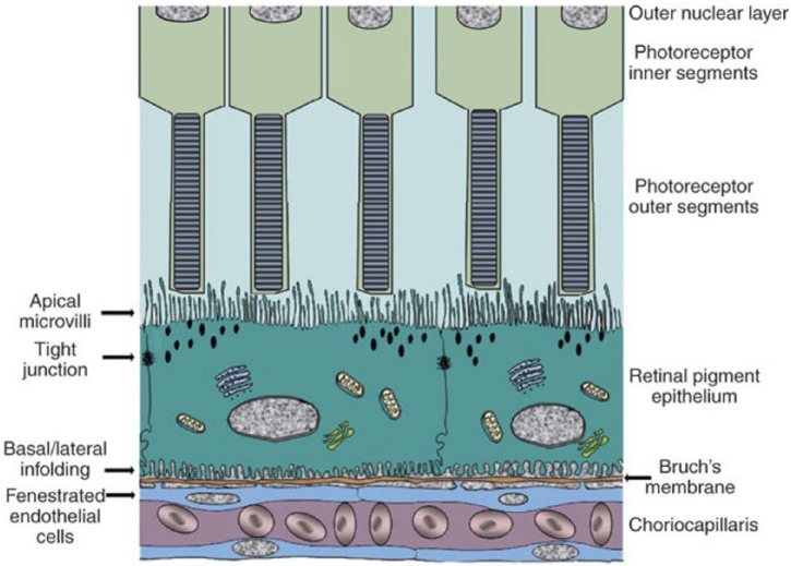
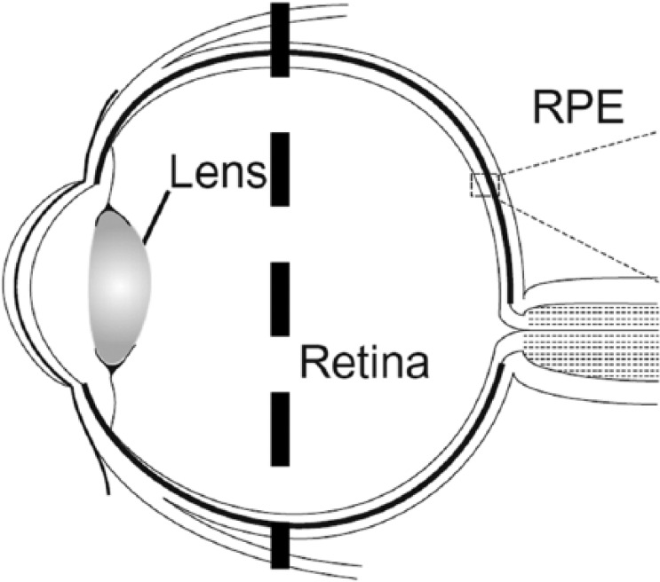
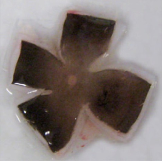
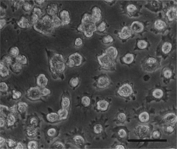
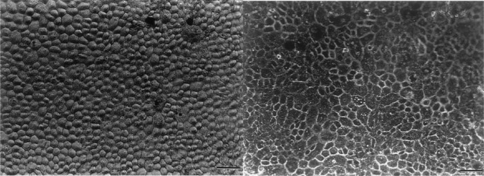
The retinal pigment epithelium is an important part of the vertebrate eye, particularly in studying the causes and possible treatment of age-related macular degeneration. The retinal pigment epithelium is difficult to access in vivo due to its location at the back of the eye, making experimentation with age-related macular degeneration treatments problematic. An alternative to in vivo experimentation is cultivating the retinal pigment epithelium in vitro, a practice that has been going on since the 1970s, providing a wide range of retinal pigment epithelial culture protocols, each producing cells and tissue of varying degrees of similarity to natural retinal pigment epithelium. The purpose of this review is to provide researchers with a ready list of retinal pigment epithelial protocols, their effects on cultured tissue, and their specific possible applications. Protocols using human and animal retinal pigment epithelium cells, derived from tissue or cell lines, are discussed, and recommendations for future researchers included.

Keywords: Retinal pigment epithelium; age-related macular degeneration; cell culture; in vitro; tissue culture.


Conflict of interest statement

Declaration of Conflicting Interests: The author(s) declared no potential conflicts of interest with respect to the research, authorship, and/or publication of this article.

Figures

Figure 1.
Figure 1.
Diagram of the outer retina, showing the retinal pigment epithelium and its location between Bruch’s membrane and the neural retina. Pigment granules, tight junctions, and monolayer formation are indicative of natural-type RPE cells.
Figure 2.
Figure 2.
Schematic of the vertebrate eye, with vertical dashed line to illustrate where incision is usually made when creating an “eyecup” from which RPE cells are harvested.
Figure 3.
Figure 3.
Dissected human fetal eyecup, with neural retina removed and RPE cells visible as dark layer on quartered tissue.
Figure 4.
Figure 4.
Porcine RPE cells in suspension after 12 min in 0.25% edetic acid during passaging. Scale bar = 50 µm.
Figure 5.
Figure 5.
Cultured porcine RPE cells, grown on a pig lens capsule (top) and a hydrogel (bottom) respectively, as found in Singh et al. Scale bar = 50 µm.

References

    1. El-Beltagy AEFBM. Light and electron microscopic studies on the pigmented epithelium and photoreceptors of the retina of common buzzard (Buteo buteo). Tissue Cell 2015; 47: 78–85. - PubMed
    1. Pequignot MO, Provost AC, Salle S, et al. The retinal pigment epithelium undergoes massive apoptosis during early differentiation and pigmentation of the optic cup. Mol Vis 2011; 17: 989–996. - PMC - PubMed
    1. Luo C, Chen M, Xu H. Complement gene expression and regulation in mouse retina and retinal pigment epithelium/choroid. Mol Vis 2011; 17: 1588–1597. - PMC - PubMed
    1. Sonoda S, Spee C, Barron E, et al. A protocol for the culture and differentiation of highly polarized human retinal pigment epithelial cells. Nat Protoc 2009; 4(5): 662–673. - PMC - PubMed
    1. Kay P, Yang YC, Paraoan L. Directional protein secretion by the retinal pigment epithelium: roles in retinal health and the development of age-related macular degeneration. J Cell Mol Med 2013; 17(7): 833–843. - PMC - PubMed