Review
. 2015 Aug 18:12:13-20.
doi: 10.2142/biophysico.12.0_13. eCollection 2015.

Three-dimensional vertex model for simulating multicellular morphogenesis

Affiliations
Review

Three-dimensional vertex model for simulating multicellular morphogenesis

Satoru Okuda et al. Biophys Physicobiol. .

Abstract

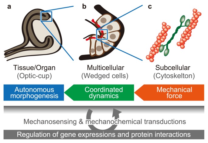
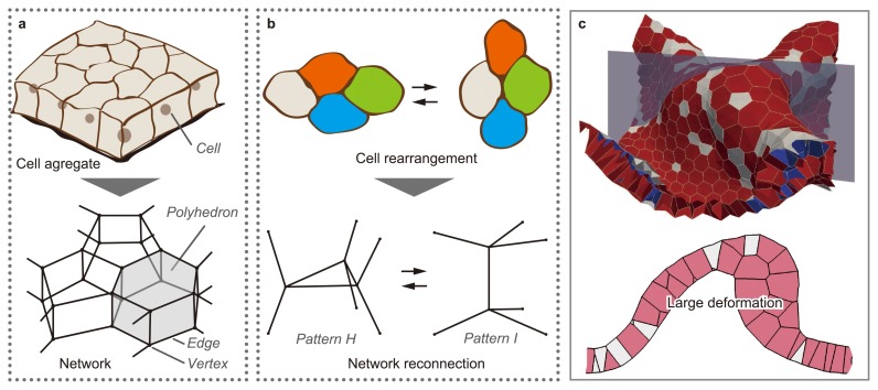
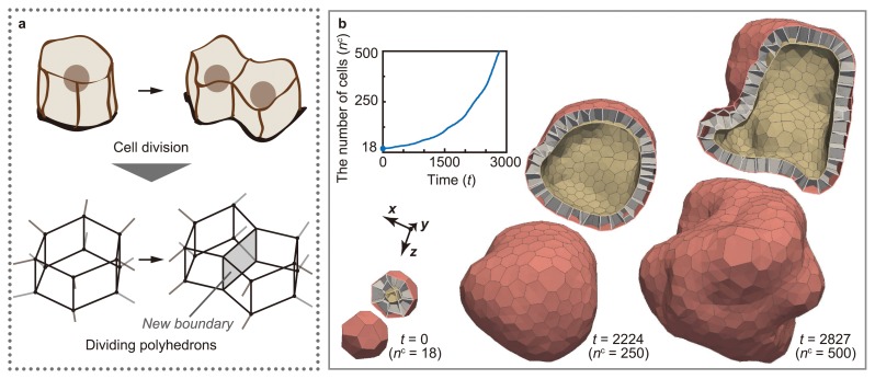
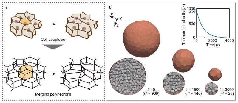
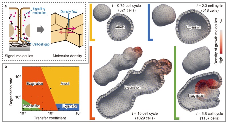
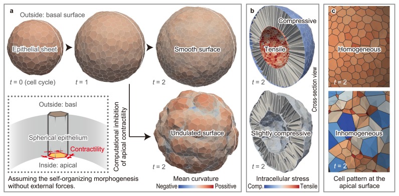
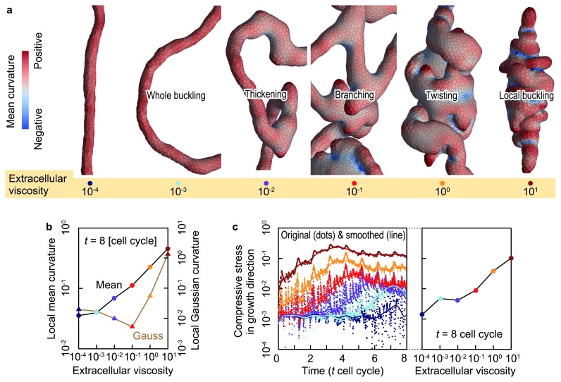
During morphogenesis, various cellular activities are spatiotemporally coordinated on the protein regulatory background to construct the complicated, three-dimensional (3D) structures of organs. Computational simulations using 3D vertex models have been the focus of efforts to approach the mechanisms underlying 3D multicellular constructions, such as dynamics of the 3D monolayer or multilayer cell sheet like epithelia as well as the 3D compacted cell aggregate, including dynamic changes in layer structures. 3D vertex models enable the quantitative simulation of multicellular morphogenesis on the basis of single-cell mechanics, with complete control of various cellular activities such as cell contraction, growth, rearrangement, division, and death. This review describes the general use of the 3D vertex model, along with its applications to several simplified problems of developmental phenomena.

Keywords: 3D vertex model; biomechanics; computational simulation; developmental biology; reversible network reconnection.


Figures

Figure 1
Figure 1
Mechanical effects of cellular activities on multicellular morphogenesis. Multicellular morphogenesis can be analyzed on several scales, such as (a) the tissue and organ scale, (b) the cell population scale, and (c) the subcellular scale. Mechanical forces generated by cellular activities on the subcellular scale cause cell–cell mechanical interactions on the cell populations scale, which drive morphogenesis on the tissue and organ scale.
Figure 2
Figure 2
Expression of cellular structure, dynamics, and rearrangement in the 3D vertex model. (a) In the 3D vertex model, the shapes of individual cells are expressed by polyhedrons, and the structure of a cell aggregate is expressed as a single network composed of the vertices and edges of these polyhedrons. (b) Cell rearrangement is expressed by reconnecting local patterns of the network according to the topological rule. (c) The model successfully simulates the large deformations of a multicellular sheet, where rearrangements of cells with changes in the layer structure are observed (Modified from [12]).
Figure 3
Figure 3
Expression of cell division in 3D vertex model. (a) Cell division is expressed by dividing polyhedrons. (b) The model of cell division successfully expresses the increase in the number of cells while regulating the timing, direction, and symmetry of cell divisions (modified from [13]).
Figure 4
Figure 4
Expression of cell death in 3D vertex model. (a) Cell death is expressed by merging polyhedrons. (b) The model of cell death successfully expresses the decrease in the number of cells, while regulating the timing of cell death. (refer to [14]).
Figure 5
Figure 5
Coupling intercellular molecular signaling with 3D multicellular deformation. (a) Intercellular molecular signaling is expressed by molecular transport between neighboring polyhedrons. (b) By simulating signal-dependent epithelial growth, we found various types of multicellular morphogenesis, such as arrest, expansion, invagination, and evagination (modified from [15]).
Figure 6
Figure 6
Effects of apical constriction on epithelial curvature. (a) Apical contractility plays a role in the maintenance of the smooth curvature of epithelial sheets amid cell division-induced disturbances. (b) Apical contractility keeps the apical surface tensile under the compressive conditions induced by cell growth. (c) The geometric cell pattern of the apical surface varies according to the presence or absence of apical contractility (modified from [24]).
Figure 7
Figure 7
The effects of extracellular viscosity on the growing epithelial tube. (a) Enlarged shapes of epithelial vesicles at several extracellular viscosities. Epithelial vesicles are colored by the local mean curvature. (b) The spatial average of the mean and Gaussian curvatures of local epithelial surfaces as a function of the extracellular viscosity. (c) Compressive forces within the epithelial vesicles in the growth direction as a function of time (left) and viscous friction weight (right). Dots and line show original and smoothed data, respectively (modified from [6]).

References

    1. Odell GM, Oster G, Alberch P.&Burnside B. The mechanical basis of morphogenesis. I. Epithelial folding and invagination. Dev Biol. 1981;85:446–462. - PubMed
    1. Honda H, Ogita Y, Higuchi S.&Kani K. Cell movements in a living mammalian tissue: long-term observation of individual cells in wounded corneal endothelia of cats. J Morphol. 1982;174:25–39. - PubMed
    1. Fletcher AG, Osborne JM, Maini PK.&Gavaghan DJ. Implementing vertex dynamics models of cell populations in biology within a consistent computational framework. Prog Biophys Mol Biol. 2013;113:299–326. - PubMed
    1. Fletcher AG, Osterfield M, Baker RE.&Shvartsman SY. Vertex models of epithelial morphogenesis. Biophys J. 2014;106:2291–2304. - PMC - PubMed
    1. Honda H.&Nagai T. Cell models lead to understanding of multi-cellular morphogenesis consisting of successive self-construction of cells. J Biochem. 2015;157:129–136. - PubMed

LinkOut - more resources