Review
. 2015 Dec 11:12:121-9.
doi: 10.2142/biophysico.12.0_121. eCollection 2015.

Microbial rhodopsins: wide distribution, rich diversity and great potential

Affiliations
Review

Microbial rhodopsins: wide distribution, rich diversity and great potential

Marie Kurihara et al. Biophys Physicobiol. .

Abstract

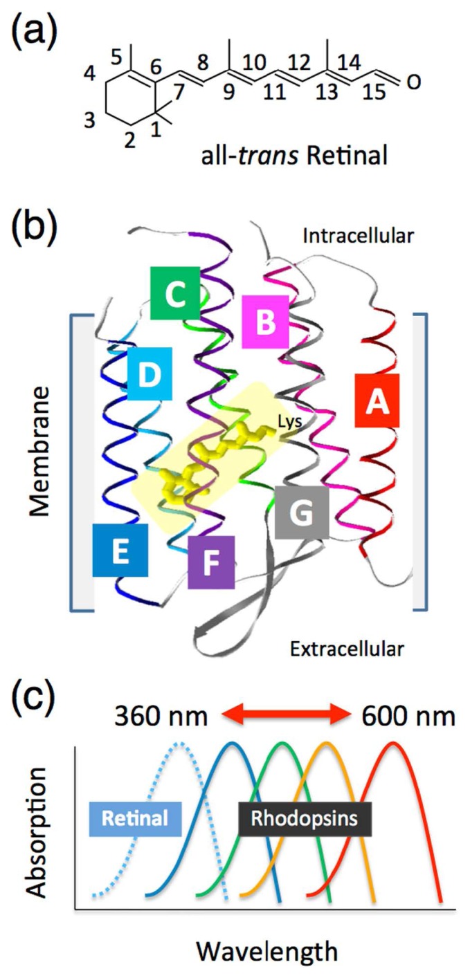
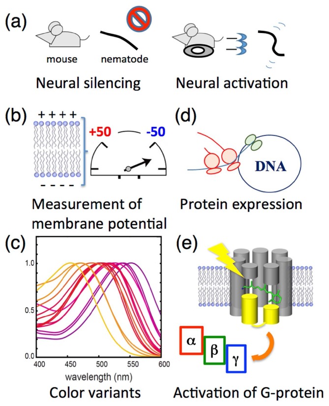
One of the major topics in biophysics and physicobiology is to understand and utilize biological functions using various advanced techniques. Taking advantage of the photoreactivity of the seven-transmembrane rhodopsin protein family has been actively investigated by a variety of methods. Rhodopsins serve as models for membrane-embedded proteins, for photoactive proteins and as a fundamental tool for optogenetics, a new technology to control biological activity with light. In this review, we summarize progress of microbial rhodopsin research from the viewpoint of distribution, diversity and potential.

Keywords: retinal; rhodopsin; visible light; vitamin-A; π-conjugation.


Figures

Figure 1
Figure 1
Introduction of the photoactive rhodopsin molecule. (a) Chemical structure of the retinal chromophore with numbering of the carbons. (b) Crystal structure of a typical type-1 microbial rhodopsin (bacteriorhodopsin, BR: PDB code, 1C3W) [71]. The retinal chromophore (yellow) that is covalently attached to the opsin via a specific lysine residue is surrounded by seven-transmembrane α-helices (A, B, C, D, E, F and G). (c) Schematic of the opsin-induced spectral shift of the retinal chromophore, named the “opsin-shift”. The absorption spectra from the retinal chromophore colored yellow in solution are greatly affected by interaction with a variety of opsins.
Figure 2
Figure 2
The classical four microbial rhodopsins from the archaeon Halobacterium salinarum. The membrane of H. salinarum contains four rhodopsins, bacteriorhodopsin (BR), halorhodopsin (HR), sensory rhodopsin-I (SRI) and sensory rhodopsin-II (SRII, also called phoborhodopsin, pR). BR and HR work as a light-driven proton pump and a halide ion pump, respectively, while SRI and SRII work as photo-sensors, and form signaling complexes with their cognate transducer proteins, HtrI and HtrII, respectively, in the membrane. Light signals are transmitted from the SRI-HtrI and SRII-HtrII complexes to a cytoplasmic two-component signal transduction cascade that consists of the adaptor protein CheW, the kinase CheA and the response regulator CheY, which regulates the rotational direction of the flagellar motor, resulting in attractant or repellent phototaxis responses. Adaptation is also essential for the detection of temporal changes of stimuli and high sensitivity to stimuli over a wide dynamic range. Covalent modifications of Htrs (i.e., methylation and demethylation), which are involved in adaptation, are illustrated as “CH3”. “P” indicates the phosphate functional group. Using those signaling systems, cells move toward longer wavelengths of light (λ>520 nm) where BR and HR work to produce ATP, while they avoid shorter wavelengths of light (λ<520 nm), which contain harmful near-UV.
Figure 3
Figure 3
The wide distribution of microbial rhodopsins in all three domains of life. By 1999, type-1 rhodopsins have only been described in halophilic archaea. The four classical rhodopsins (BR, HR, SRI and SRII) are shown as boxes with white letters. After 1999, environmental genomics revealed that microbial rhodopsins are broadly distributed among bacteria (green) and eukarya (blue) in addition to archaea (red).
Figure 4
Figure 4
Multitalented microbial rhodopsins. The type-1 microbial rhodopsins are roughly categorized as ion transporters (i.e., cation pump, anion pump and ion channel) and as sensors. (a) For ion transporters, the number of substrates (X, Y and Z) greatly increased in the early 21st century. (b) For sensors and coenzymes, the photosensory function was also extended from only the regulation of kinases to the control of cyclases, transcriptional regulation and depolarization of the plasma membrane that accounts for the phototactic behaviors of the prokaryote Chlamydomonas reinhardtii.
Figure 5
Figure 5
Optogenetic applications. (a) The photo-activation of ion transporting rhodopsins induces a hyperpolarization used for neural silencing and a depolarization used for neural activation both in vivo and in vitro. (b) The genetically modified H+ pump AR3 is utilized to measure the membrane potential in vivo. (c) The color variants of ion transporting rhodopsins allow optogenetics control in a wide range of wavelengths of light. (d) One of the sensory rhodopsins, Anabaena sensory rhodopsin (ASR), can be utilized as a tool for arbitrary protein expression in vivo regulated by visible light. (e) Chimeric proteins of Gloeobacter rhodopsin (GR) and bovine visual rhodopsin (Rh) are utilized to activate the trimeric G-protein.

References

    1. Ernst OP, Lodowski DT, Elstner M, Hegemenn P, Brown LS.&Kandori H. Microbial and animal rhodopsins: structures, functions, and molecular mechanisms. Chem Rev. 2014;114:126–163. - PMC - PubMed
    1. Sudo Y. CRC Handbook of Org Photochem Photobiol. 3rd eds. CRC Press; Boca Raton: 2012. Transport and sensory rhodopsins in microorganisms; pp. 1173–1193.
    1. Spudich JL, Yang CS, Jung KH.&Spudich EN. Retinylidene proteins: structures and functions from archaea to humans. Annu Rev Cell Dev Biol. 2000;16:365–392. - PubMed
    1. Brown LS. Eubacterial rhodopsins—unique photosensors and diverse ion pumps. Biochim Biophys Acta. 2014;1837:553–561. - PubMed
    1. Brown LS.&Jung KH. Bacteriorhodopsin-like proteins of eubacteria and fungi: the extent of conservation of the haloarchaeal proton-pumping mechanism. Photochem Photobiol Sci. 2006;5:538–546. - PubMed