. 2016 Aug 3;2(8):e1501429.
doi: 10.1126/sciadv.1501429. eCollection 2016 Aug.

Microfluidic device for the formation of optically excitable, three-dimensional, compartmentalized motor units

Affiliations

Microfluidic device for the formation of optically excitable, three-dimensional, compartmentalized motor units

Sebastien G M Uzel et al. Sci Adv. .

Abstract

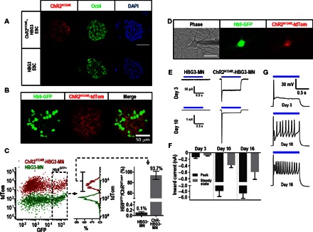
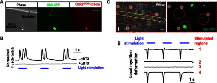
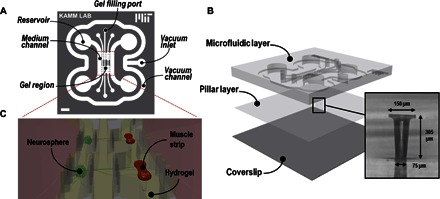
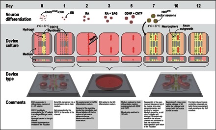
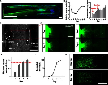
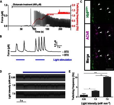
Motor units are the fundamental elements responsible for muscle movement. They are formed by lower motor neurons and their muscle targets, synapsed via neuromuscular junctions (NMJs). The loss of NMJs in neurodegenerative disorders (such as amyotrophic lateral sclerosis or spinal muscle atrophy) or as a result of traumatic injuries affects millions of lives each year. Developing in vitro assays that closely recapitulate the physiology of neuromuscular tissues is crucial to understand the formation and maturation of NMJs, as well as to help unravel the mechanisms leading to their degeneration and repair. We present a microfluidic platform designed to coculture myoblast-derived muscle strips and motor neurons differentiated from mouse embryonic stem cells (ESCs) within a three-dimensional (3D) hydrogel. The device geometry mimics the spinal cord-limb physical separation by compartmentalizing the two cell types, which also facilitates the observation of 3D neurite outgrowth and remote muscle innervation. Moreover, the use of compliant pillars as anchors for muscle strips provides a quantitative functional readout of force generation. Finally, photosensitizing the ESC provides a pool of source cells that can be differentiated into optically excitable motor neurons, allowing for spatiodynamic, versatile, and noninvasive in vitro control of the motor units.

Keywords: Microfluidics; neuromuscular junctions; optogenetics; tissue engineering.


Figures

Fig. 1
Fig. 1. ChR integration via homologous recombination results in stable expression in ESC and ES-derived MNs and proper light-driven neuronal stimulation.
(A) Membrane-bound expression of tdTomato-tagged ChR2 is observed in transfected ES colonies. Immunostaining for Oct4 expression confirms the pluripotent nature of the transformed cells. Scale bar, 50 μm. DAPI, 4′,6-diamidino-2-phenylindole. (B) Confocal image of a ChR-HBG3–derived neurosphere on day 7 after RA and SAG treatment, showing persistent expression of ChR. (C) FACS data comparing tdTomato::ChR expression of parental (HBG3-MN) and ChR-expressing (ChR-HBG3-MN) cells dissociated from day 7 neurospheres, demonstrating robust expression and minimum silencing after reaching the MN lineage. (D) Dissociated Hb9GFP+/ChRtdTom+ MN plated on a monolayer of cortical glial feeder cells assuming proper neuronal morphology on day 3. The phase contrast image features the patching electrode. Scale bar, 50 μm. (E) Representative trace displaying inward current upon optical stimulation (blue bar) on days 3 and 10 on HBG3-MN and ChRH134R-HBG3-MN. (F) Peak and steady-state inward currents on days 3, 10, and 16 in ChR-HBG3-MN (n = 10). Error bars, SD. (G) Representative current-clamp recordings upon prolonged 1-s optical stimulation displaying AP elicitation on days 3, 10, and 16.
Fig. 2
Fig. 2. ChR-HBG3-MN form functional NMJs in vitro in adherent cultures.
(A) Dissociated Hb9GFP+/ChRtdTom+ MN forming initial contact with a C2C12-derived myotube after 1 day of coculture. Scale bar, 50 μm. (B) Muscle contraction observed upon optical stimulation (blue bar) of the ChR-HBG3-MN. The contractions were inhibited after incubation with αBTX. (C) Local optogenetic excitation of neuron-muscle coculture: (i) phase contrast and epifluorescence images of ChR-HBG3-MN and muscle cells. The myotube of interest is outlined in yellow, and the stimulated regions are outlined by the red dashed line. Scale bar, 100 μm. (ii) Muscle twitch [outlined in yellow in (i)] as light stimulation (blue bars) is applied to various regions outlined in red: full field of view (1), muscle only (2), noninnervating MN cluster (3), and innervating cluster (4).
Fig. 3
Fig. 3. Microfluidic design and assembly.
(A) The microfluidic design features three parallel gel regions accessible by six gel filling ports and flanked by two medium channels connected to four medium reservoirs. A surrounding vacuum channel allows for temporary bonding. Scale bar, 2 mm. (B) The platform is composed of a top microfluidic layer assembled on top of a PDMS membrane featuring two sets of two capped pillars (inset), itself bonded to a coverslip. (C) Schematic displaying the final coculture arrangement: embedded in a hydrogel, muscle bundles that are wrapped around and exerted force to the pillars are innervated by neurospheres located in the opposite gel chamber separated by a 1-mm-wide gel region.
Fig. 4
Fig. 4. Framework for the microfluidic neuromuscular coculture.
(Row 1) Schematic showing the differentiation process of the ESCs into MNs following a previously published protocol (37). (Row 2) Schematics displaying the top and front views of the tissue in the microfluidic platform. (Row 3) 3D computer-aided drafting illustrations showing the version of the platform used at the corresponding days. CNTF, ciliary neurotrophic factor; HS, horse serum.
Fig. 5
Fig. 5. Muscle differentiation and neuromuscular tissue formation.
(A) Immunostaining of the myogenic marker α-actinin (green) and DAPI (blue), demonstrating proper muscle differentiation and formation of sarcomeric striations (examples of striations are indicated by arrowheads in the inset). Scale bars, 50 μm. (B) Muscle bundle width relative to the width at day 0. (C) Passive force generation over the course of 16 days. (D) Representative image of a neuron-muscle coculture in the microfluidic device on day 1 of coculture. Scale bar, 500 μm. (E) Neurite extension over 4 days of coculture. Scale bar, 250 μm. (F) Absolute maximum neurite outgrowth in millimeters over the first 4 days of culture. For comparison, the red dashed line represents the average initial distance between neurospheres and muscle bundles (the shaded area in between the dotted line indicates the SEM). (G) Percentage of muscle bundles contacted by at least one neurite over the course of 4 days. (H) Confocal 3D reconstruction of neurites in the bridge gel region after 1 day of coculture. Scale bar, 200 μm. All error bars, SEM.
Fig. 6
Fig. 6. Activation of NMJs within the microfluidic device.
(A) Application of glutamate to the medium results in a delayed stimulation of the muscle, leading to the initiation of muscle twitching with force (left y axis) at an increasing frequency (right y axis) as glutamate diffuses within the neurospheres. (B) Force generated by the muscle bundle upon illumination of the ChR2H134R-HBG3-MN neurospheres on day 15. Application of αBTX inhibited the contractions. (C) Colocalization of incoming motor axons and clusters of AChR indicative of the presence of NMJ. Scale bar, 100 μm. (D) Kymographs of the pillar displacement on day 16 for three stimulation light intensities. (E) Muscle-twitching frequencies as a function of light intensity. *P < 0.05, **P < 0.001, ***P < 0.0005.

References

    1. Olesen J., Gustavsson A., Svensson M., Wittchen H.-U., Jönsson B. CDBE2010 study group European Brain Council , The economic cost of brain disorders in Europe. Eur. J. Neurol. 19, 155–162 (2012). - PubMed
    1. Dale H. H., Feldberg W., Vogt M., Release of acetylcholine at voluntary motor nerve endings. J. Physiol. 86, 353–380 (1936). - PMC - PubMed
    1. Fatt P., Katz B., Spontaneous subthreshold activity at motor nerve endings. J. Physiol. 117, 109–128 (1952). - PMC - PubMed
    1. Sanes J. R., Lichtman J. W., Development of the vertebrate neuromuscular junction. Annu. Rev. Neurosci. 22, 389–442 (1999). - PubMed
    1. Cao G., Ko C.-P., Schwann cell-derived factors modulate synaptic activities at developing neuromuscular synapses. J. Neurosci. 27, 6712–6722 (2007). - PMC - PubMed

Publication types

MeSH terms

Substances