. 2016 Aug 3;2(8):e1600418.
doi: 10.1126/sciadv.1600418. eCollection 2016 Aug.

Battery-free, stretchable optoelectronic systems for wireless optical characterization of the skin

Affiliations

Battery-free, stretchable optoelectronic systems for wireless optical characterization of the skin

Jeonghyun Kim et al. Sci Adv. .

Abstract

Recent advances in materials, mechanics, and electronic device design are rapidly establishing the foundations for health monitoring technologies that have "skin-like" properties, with options in chronic (weeks) integration with the epidermis. The resulting capabilities in physiological sensing greatly exceed those possible with conventional hard electronic systems, such as those found in wrist-mounted wearables, because of the intimate skin interface. However, most examples of such emerging classes of devices require batteries and/or hard-wired connections to enable operation. The work reported here introduces active optoelectronic systems that function without batteries and in an entirely wireless mode, with examples in thin, stretchable platforms designed for multiwavelength optical characterization of the skin. Magnetic inductive coupling and near-field communication (NFC) schemes deliver power to multicolored light-emitting diodes and extract digital data from integrated photodetectors in ways that are compatible with standard NFC-enabled platforms, such as smartphones and tablet computers. Examples in the monitoring of heart rate and temporal dynamics of arterial blood flow, in quantifying tissue oxygenation and ultraviolet dosimetry, and in performing four-color spectroscopic evaluation of the skin demonstrate the versatility of these concepts. The results have potential relevance in both hospital care and at-home diagnostics.

Keywords: Battery-free; NFC; bio-integrated; healthcare; near-field communication; optical; optoelectronics; skin; stretchable; wireless.


Figures
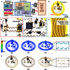
Fig. 1
Fig. 1. Wireless epidermal optoelectronics.
(A) Block diagram of the system, including its NFC wireless components for wireless power transfer and data communication. An external set of reader electronics delivers power to the device through magnetic inductive coupling. This power activates the optoelectronic components, the analog/digital (A/D) converter, and the NFC hardware for wireless transmission of the output of the photodetector back to the reader, where it is recorded for further processing. (B) Image of a complete device configured to measure heart rate. (C to E) Magnified sections of (B). The system includes an IR LED and a photodetector (C), an amplifier and resistors for conditioning (D), and an inductive coil (E). (F) FEA at the system level reveals the displacement and strain distributions for uniaxial strains up to 30%. (G) Corresponding images of the device. The inset highlights a highly deformed region for both modeling and experiment. The system functions properly even at the highest strains illustrated here.
Fig. 2
Fig. 2. Wireless epidermal optoelectronic system with a single LED and photodetector designed for heart rate and MAP tracking.
(A) Exploded-view illustration of the device construction. (B) Image of a device mounted on the skin while deformed by pinching. (C) Image of the device during wireless operation with a smartphone, for both power deliver and data communication. (D) Wireless measurement results during recording on the forearm. (E) Magnified view of the red dashed box in (D), with an inverted y-axis scale, for ease of viewing. The systolic peak and the dicrotic notch, evident in these data, correspond to the maximum pressure generated during the systolic ejection and to the closing of the aorta, respectively. The MAP relates to the area under the pulse waveform. (F) Fourier transform of the signal in (D). The graphs show three harmonics, the first one of which corresponds to the beat rate (~1.5 Hz or 90 beats per minute).
Fig. 3
Fig. 3. Wireless epidermal optoelectronic system with two pulsed LEDs and a single photodetector to monitor peripheral vascular disease.
(A) Image of an epidermal wireless oximeter that includes a red LED, an IR LED, a photodiode, and associated electronics all in a stretchable configuration mounted on a soft, black textile substrate coated with a low-modulus silicone elastomer. (B) Schematic illustration of the circuit of the device. An astable oscillator switches current between the two LEDs to allow time-multiplexed measurement of both wavelengths with a single photodetector. The R1C1 and R2C2 tanks set the frequency of the oscillator. GND, ground. (C) Images of the device operating during activation of the red LED (top) and the infrared LED (bottom). (D) Image of the device mounted on the forearm. (Inset) Schematic illustration of the operating principle. (E) Functional demonstration in a procedure that involves transient vein occlusion (gray box in the graph). An inflating cuff on participant’s bicep temporarily occludes venous blood flow set to a pressure slightly below the arterial pressure (50 mmHg). (F) Magnified view of the red dashed box in (E). (G and H) Measurements obtained by a commercial oximeter and an epidermal device, simultaneously recorded from adjacent regions of the forearm. NIRS, NIR spectroscopy.
Fig. 4
Fig. 4. Wireless epidermal optoelectronic system with two pulsed LEDs, a single photodetector, and a colorimetric responsive material designed for UV dosimetry.
(A and B) Images of the device, which includes a red LED, an IR LED, and a photodiode, all encapsulated with thin, stretchable silicone film doped with a dye that changes color upon exposure to UV light. The two LEDs switch at different frequencies, through the use of an astable oscillator, to enable time-multiplexed readout with a single photodetector. (C) Optical transmittance of the UV-sensitive layer at different UV exposure dose levels, with corresponding images (the material changes from blue to transparent). (D) Sketch illustrating the operating principle. The LEDs emit light laterally through the UV-responsive layer. The use of one red and one IR LED enables differential measurement of the transmitted light. (E and F) Measurements at varying times of exposure to UV light from a solar simulator. The changes in transmission occur mainly at the red wavelength (640 nm), consistent with spectroscopic characterization of the material in (C). The ratio of the two signals is independent of both the power and the bias conditions of the LEDs. a.u., arbitrary units.
Fig. 5
Fig. 5. Wireless epidermal optoelectronic system with four pulsed LEDs and a single photodetector for spectrophotometric characterization.
(A) Image of a wireless spectrometer that includes four pulsed LEDs, each with a different color (red, IR, orange, and yellow). There are two switching LED pairs in the device (red-IR and yellow-orange). Two astable oscillators control the switching at four different frequencies to distinguish each signal. (B) Images of the device operating while the LEDs are activated. (C) Image of a device operating while submerged in water captured at a moment during activation of red and yellow LEDs. (D) Reflectance measurement of three different colored apples. The vertical lines denote the wavelength of each LED light. (E) Wireless measurement of three different colored apples. (F) Calculated reflectance from measurement data of apples with different colors. (G) Images of subjects with different skin colors. (H) Wireless measurement data of skins with varying colors. (I) Calculated reflectance from the measurement data of skins with different colors.

References

    1. Steinhubl S. R., Muse E. D., Topol E. J., The emerging field of mobile health. Sci. Transl. Med. 7, 283rv3 (2015). - PMC - PubMed
    1. Monton E., Hernandez J. F., Blasco J. M., Hervé T., Micallef J., Grech I., Brincat A., Traver V., Body area network for wireless patient monitoring. IET Commun. 2, 215–222 (2008).
    1. Boppart S. A., Richards-Kortum R., Point-of-care and point-of-procedure optical imaging technologies for primary care and global health. Sci. Transl. Med. 6, 253rv2 (2014). - PMC - PubMed
    1. Otto C., Milenković A., Sanders C., Jovanov E., System architecture of a wireless body area sensor network for ubiquitous health monitoring. J. Mobile Multimedia 1, 307–326 (2006).
    1. Jovanov E., Milenkovic A., Otto C., de Groen P. C., A wireless body area network of intelligent motion sensors for computer assisted physical rehabilitation. J. Neuroeng. Rehabil. 2, 6 (2005). - PMC - PubMed

Publication types