. 2016 Aug 5;12(8):e1005035.
doi: 10.1371/journal.pcbi.1005035. eCollection 2016 Aug.

Strain Mediated Adaptation Is Key for Myosin Mechanochemistry: Discovering General Rules for Motor Activity

Affiliations

Strain Mediated Adaptation Is Key for Myosin Mechanochemistry: Discovering General Rules for Motor Activity

Biman Jana et al. PLoS Comput Biol. .

Abstract

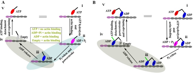
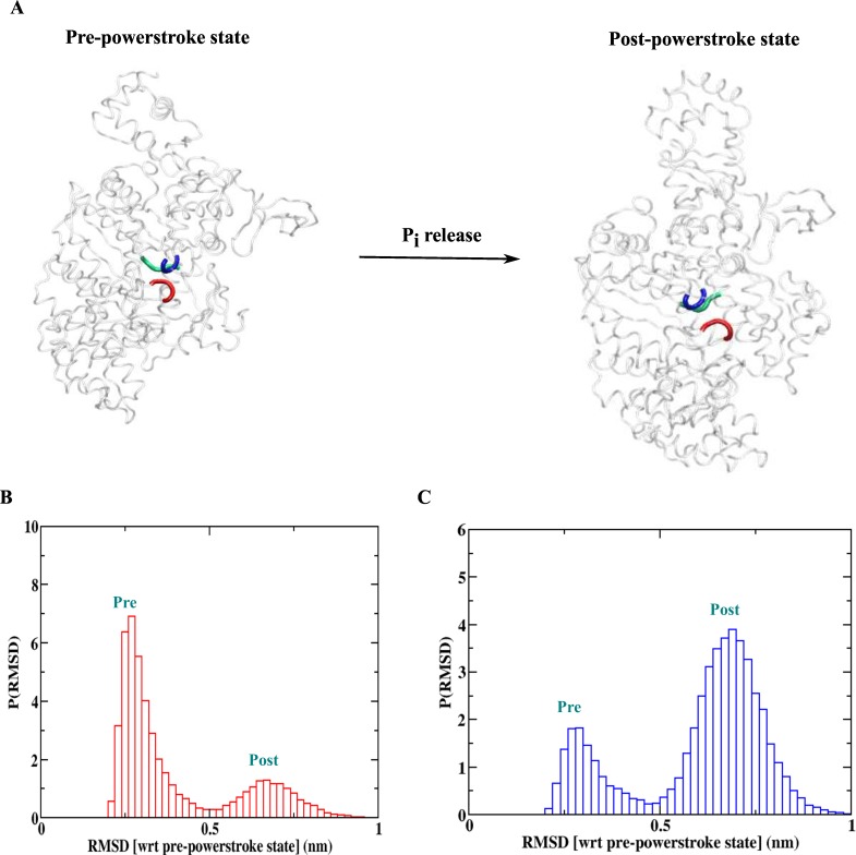
A structure-based model of myosin motor is built in the same spirit of our early work for kinesin-1 and Ncd towards physical understanding of its mechanochemical cycle. We find a structural adaptation of the motor head domain in post-powerstroke state that signals faster ADP release from it compared to the same from the motor head in the pre-powerstroke state. For dimeric myosin, an additional forward strain on the trailing head, originating from the postponed powerstroke state of the leading head in the waiting state of myosin, further increases the rate of ADP release. This coordination between the two heads is the essence of the processivity of the cycle. Our model provides a structural description of the powerstroke step of the cycle as an allosteric transition of the converter domain in response to the Pi release. Additionally, the variation in structural elements peripheral to catalytic motor domain is the deciding factor behind diverse directionalities of myosin motors (myosin V & VI). Finally, we observe that there are general rules for functional molecular motors across the different families. Allosteric structural adaptation of the catalytic motor head in different nucleotide states is crucial for mechanochemistry. Strain-mediated coordination between motor heads is essential for processivity and the variation of peripheral structural elements is essential for their diverse functionalities.


Conflict of interest statement

The authors have declared that no competing interests exist.

Figures

Fig 1
Fig 1. Functional cycle of single- and double-headed myosin.
(A) Sequence of events of the mechanochemical cycle of the single-headed myosin. ATP bound (red) head (i) binds to the actin filament followed by the hydrolysis of ATP. The arm (black line on head) of this actin bound head (ii) in ADP + Pi state (pink) has a pre-powerstroke conformation. Phosphate (Pi) release induces the powerstroke conformational change to the lever arm (iii). Next, ADP (blue) is released from the bound head while keeping the post-powerstroke lever arm conformation (iv). Finally, the empty head (gray) detaches from the actin followed by an ATP intake. The ATP dependent unbound head experiences a repriming event to its stable pre-powerstroke lever arm conformation (v). Note that, state (v) is exactly same as state (i) with an additional stepping towards left and also the nucleotide dependent actin binding affinity information in the middle. (B) The sequence of events of the functional cycle of the double-headed myosin. In state (i), head 1 is in an ATP bound state and head 2 is bound to actin in an ADP state. ATP hydrolysis and subsequent binding to actin by head 1 provide a two-head bound myosin (ii). Head 1 releases phosphate (Pi) to transform into state (iii). In contrary to a single-head myosin, here, after Pi release, head 1 cannot perform powerstroke while head 2 is still bound. The green line shows the expected lever arm conformation of head 1. It is important to note here that the two-head-bound state iii could adopt an alternative conformation with the converters of both heads in a post-powerstroke conformation while the lever arm of the leading/trailing head bends backward/forward. Next, ADP releases from head 2 (iv) and subsequently ATP binds to the empty head. Head 2 now detaches from the actin and head 1 now performs its postponed powerstroke step. Finally, head 2 also perform its spontaneous repriming event to form state (v). Note that, state (v) is exactly same as state (i) with an additional stepping towards the left.
Fig 2
Fig 2. Two important conformational states of the mechanical cycle of myosin.
(A) Pre- (PDB 2V26 [34]) and post-powerstroke (PDB 2BHK [36]) states of the myosin motor showing the motor head (MH) and the converter domains. Note the change in the conformation of the converter domain with respect to the MH in the two conformations. The competing interactions (contacts) between the MH and the converter domains, responsible for stabilizing these two states, are shown by lines. (B) Residue-residue contact map for both the conformations. The two dotted green lines are drawn to separate the contact maps of the MH and the converter domains. In the upper triangle, the post-powerstroke contact map (red) is overlaid upon the pre-powerstroke contact map (blue) to identify the exclusive converter-MH contacts in the pre-powerstroke state (shown by lines in Fig 2A). In contrast, the same data have been overlaid in opposite order (blue upon red) in the lower triangle to extract the exclusive converter-MH contacts in the post-powerstroke state (those interactions are shown by lines in Fig 2A).
Fig 3
Fig 3. Structural adaptation of the trailing head signals faster ADP release.
(A) Myosin MH domain structure showing the big (red) and small (green) subunits. (B) Nucleotide binding region of the MH domain is shown in terms of P-loop (blue), switch I (red) and switch II (green). (C) RMSD distribution (P(RMSD)) of the small subunit of the MH domain after least square fitting of the big subunit from the leading and trailing head simulations. The RMSD is calculated with respect to the pre-powerstroke MH conformation. Note the larger RMSD for the trailing head indicating substantial structural changes. (D) Distribution of distances between P-loop and switch I (P(dSWI-Ploop)) for the leading and trailing head simulations. (E) Distribution of distances between P-loop and switch II (P(dSWII-Ploop)) for the leading and trailing head simulations. (F) Distribution of distances between switch I and switch II (P(dSWI-SWII)) for the leading and trailing head simulations. A larger distance between switch I and switch II for the trailing head simulation compared to leading head signals faster ADP release.
Fig 4
Fig 4. Strain regulates the ADP release kinetics of the trailing head.
(A) A schematic representation of myosin dimer showing how Pi release can create strain on the trailing head. This strain arises due to the postponed-powerstroke waiting state of the leading head (left head). (B) Probability distribution of distances between small and big subunits of the MH domain at different level of strain on the trailing head. This distribution shifts towards the larger distances as strain increases, which leads to an increase in ADP release rate with increasing strain.
Fig 5
Fig 5. Powerstroke of the myosin motors.
(A) Powerstroke step with the release of Pi. The residues involved in the Pi mediated interactions in terms of p-loop (blue), switch I (red) and switch II (green) are shown. (B) RMSD (with respect to pre-powerstroke crystal structure) distribution for the simulation with Pi mediated interaction. The population of the pre-powerstroke ensemble is higher. (C) RMSD (with respect to pre-powerstroke crystal structure) distribution for the simulation without Pi mediated interactions. Note the inversion of the distribution with phosphate release making post-powerstroke ensemble as the dominant one.

Similar articles

Cited by

References

    1. Reedy MC. Visualizing myosin’s power stroke in muscle contraction. J Cell Sci. 2000; 113: 3551–3562 - PubMed
    1. Odronitz F, Kollmar M. Drawing the tree of eukaryotic life based on the analysis of 2, 269 manually annotated myosins from 328 species. Gen Biol.2007; 8: R196 - PMC - PubMed
    1. Goldman YE. Wag the tail: structural dynamics of actomyosin. Cell.1998; 93: 1–4. - PubMed
    1. Moore JR, Krementsova EB, Trybus KM, Warshaw DM. Myosin V exhibits a high duty cycle and large unitary displacement. J Cell Biol. 2001;155: 625–635. - PMC - PubMed
    1. De La Cruz EM, Ostap EM. Relating biochemistry and function in the myosin superfamily. Curr Opin Cell Biol. 2004;16:61–67. - PubMed

Publication types

MeSH terms