

Circadian Rhythm Regulates Development of Enamel in Mouse Mandibular First Molar
- PMID: 27494172
- PMCID: PMC4975438
- DOI: 10.1371/journal.pone.0159946
Circadian Rhythm Regulates Development of Enamel in Mouse Mandibular First Molar
Abstract
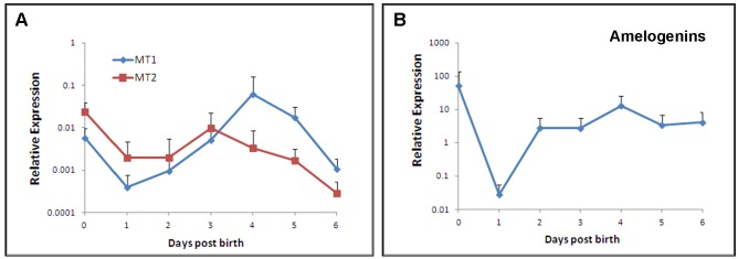
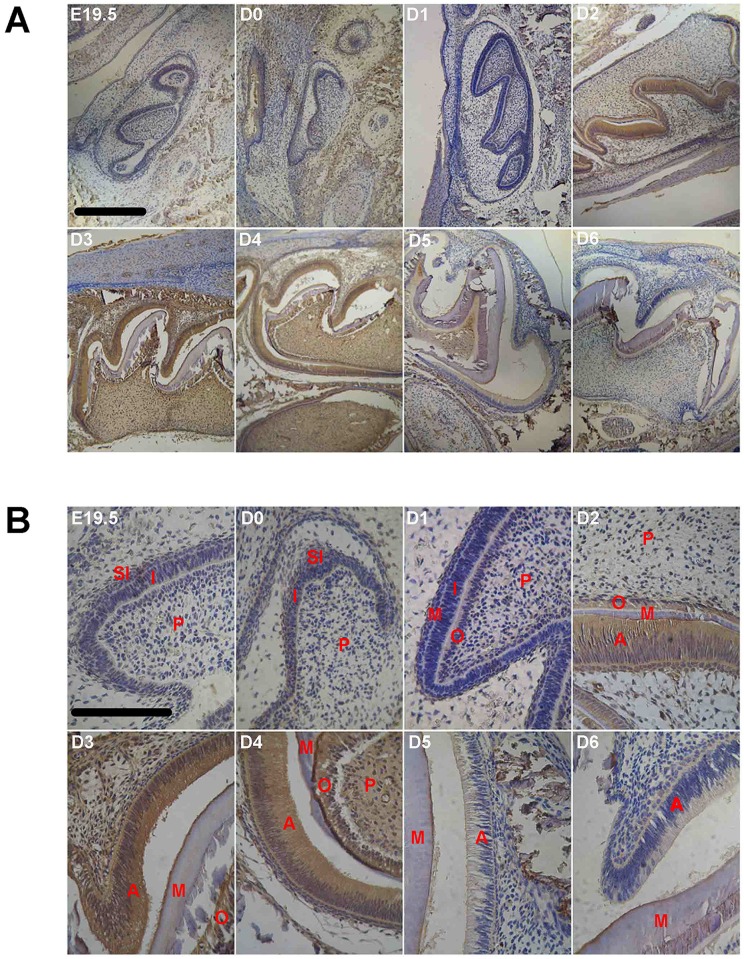
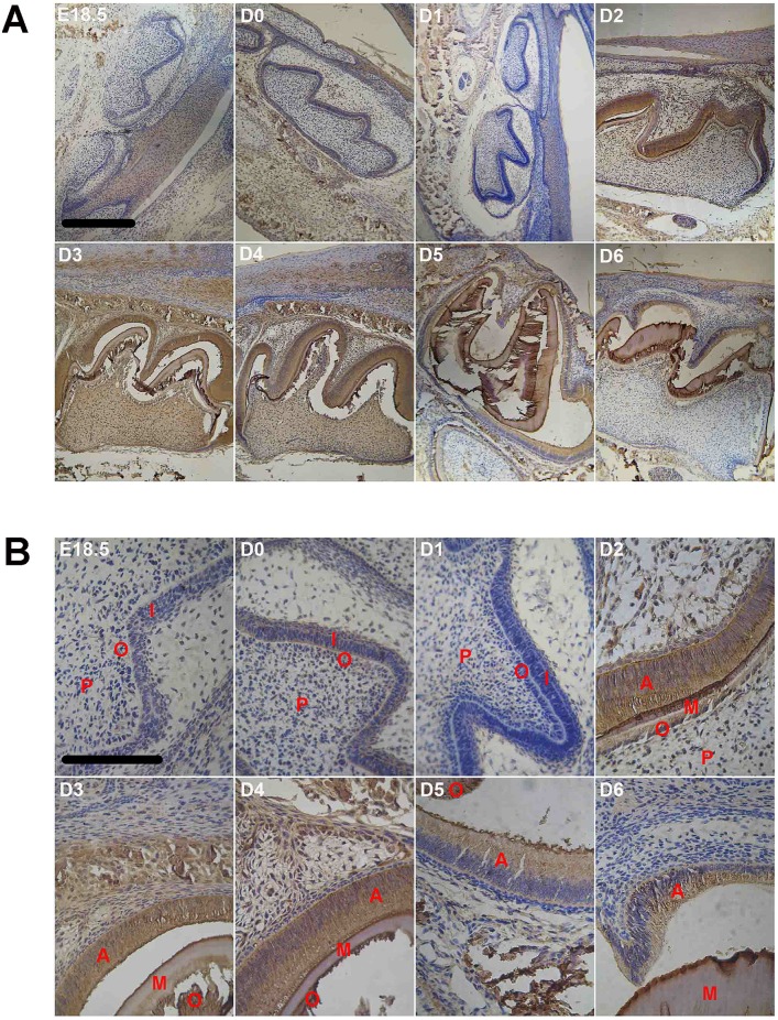
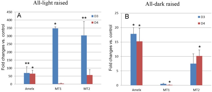
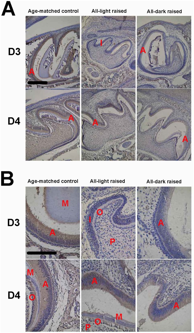
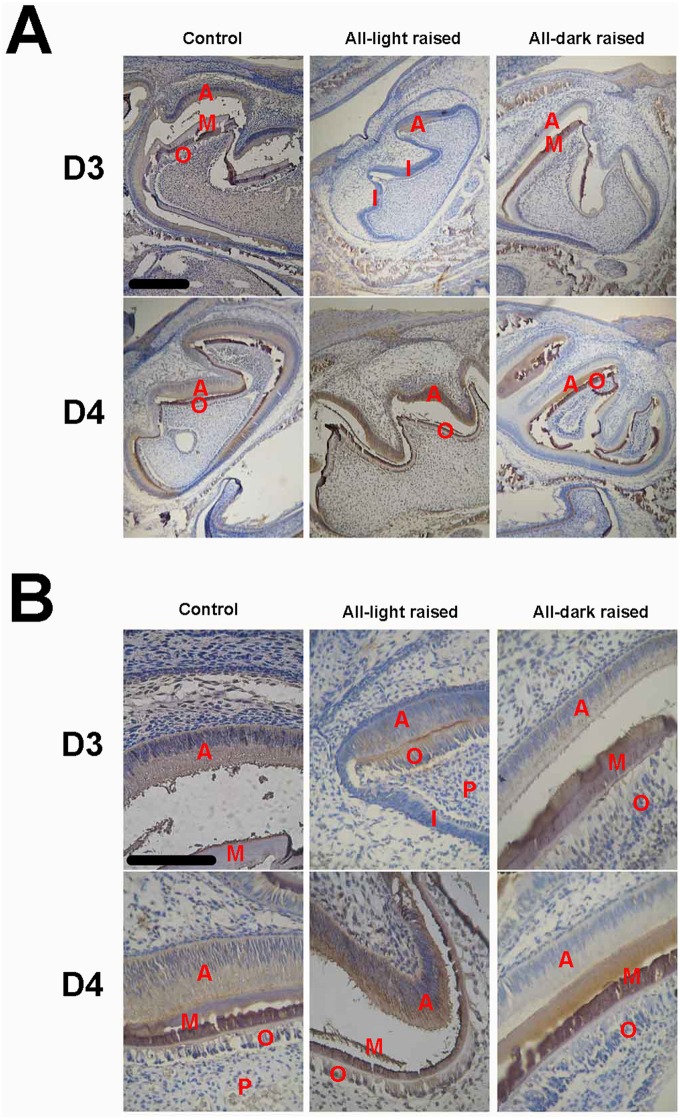
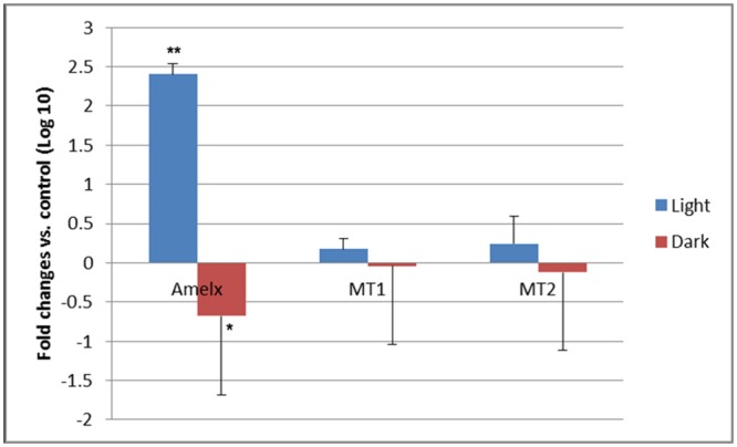
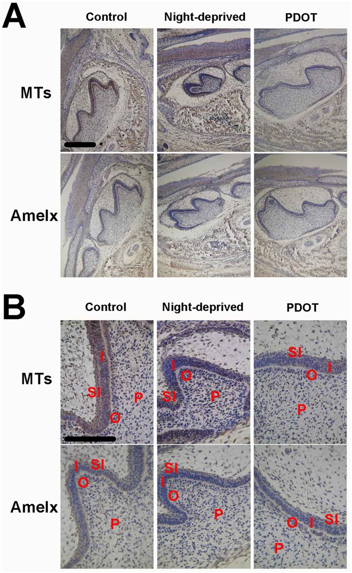
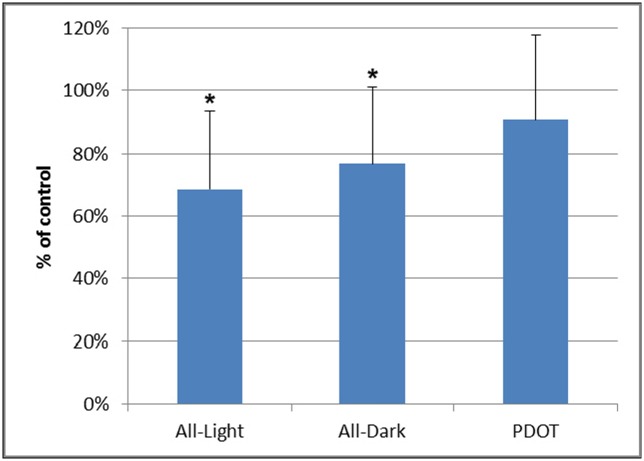
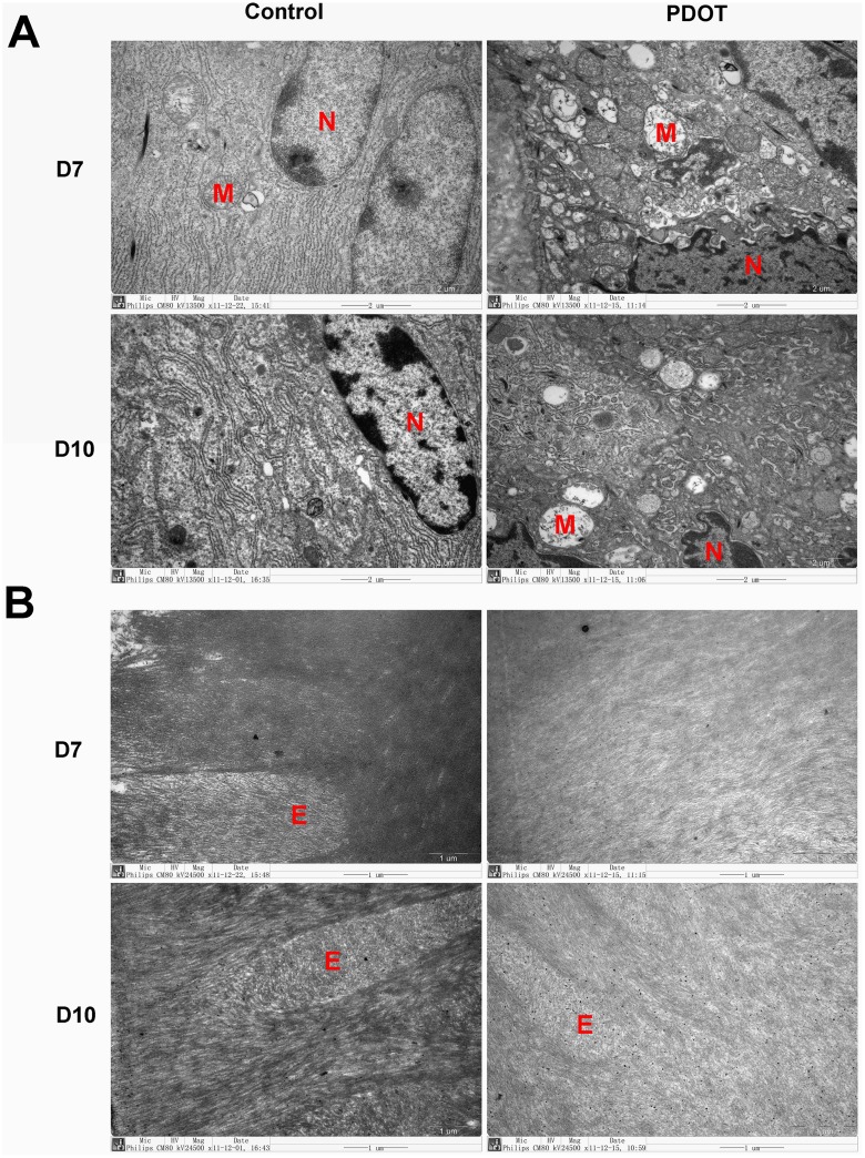
Rhythmic incremental growth lines and the presence of melatonin receptors were discovered in tooth enamel, suggesting possible role of circadian rhythm. We therefore hypothesized that circadian rhythm may regulate enamel formation through melatonin receptors. To test this hypothesis, we examined expression of melatonin receptors (MTs) and amelogenin (AMELX), a maker of enamel formation, during tooth germ development in mouse. Using qRT-PCR and immunocytochemistry, we found that mRNA and protein levels of both MTs and AMELX in normal mandibular first molar tooth germs increased gradually after birth, peaked at 3 or 4 day postnatal, and then decreased. Expression of MTs and AMELX by immunocytochemistry was significantly delayed in neonatal mice raised in all-dark or all-light environment as well as the enamel development. Furthermore, development of tooth enamel was also delayed showing significant immature histology in those animals, especially for newborn mice raised in all daylight condition. Interestingly, disruption in circadian rhythm in pregnant mice also resulted in delayed enamel development in their babies. Treatment with melatonin receptor antagonist 4P-PDOT in pregnant mice caused underexpression of MTs and AMELX associated with long-lasting deficiency in baby enamel tissue. Electromicroscopic evidence demonstrated increased necrosis and poor enamel mineralization in ameloblasts. The above results suggest that circadian rhythm is important for normal enamel development at both pre- and postnatal stages. Melatonin receptors were partly responsible for the regulation.
Conflict of interest statement
Figures
Similar articles
-
[WWP1 plays a positive role in ameloblast differentiation and enamel formation in mice].Zhonghua Kou Qiang Yi Xue Za Zhi. 2025 Jan 9;60(1):33-42. doi: 10.3760/cma.j.cn112144-20240916-00349. Zhonghua Kou Qiang Yi Xue Za Zhi. 2025. PMID: 39743363 Chinese.
-
[Expression patterns of amelogenin and enamelin in developing mouse tooth germs].Zhonghua Kou Qiang Yi Xue Za Zhi. 2012 Mar;47(3):173-6. doi: 10.3760/cma.j.issn.1002-0098.2012.03.011. Zhonghua Kou Qiang Yi Xue Za Zhi. 2012. PMID: 22800672 Chinese.
-
[Immunohistochemical localization of enamelin in developing rat tooth germ].Beijing Da Xue Xue Bao Yi Xue Ban. 2007 Feb 18;39(1):37-40. Beijing Da Xue Xue Bao Yi Xue Ban. 2007. PMID: 17304324 Chinese.
-
Essential roles of ameloblastin in maintaining ameloblast differentiation and enamel formation.Cells Tissues Organs. 2005;181(3-4):189-95. doi: 10.1159/000091380. Cells Tissues Organs. 2005. PMID: 16612084 Review.
-
The circadian clock in enamel development.Int J Oral Sci. 2024 Sep 6;16(1):56. doi: 10.1038/s41368-024-00317-9. Int J Oral Sci. 2024. PMID: 39242565 Free PMC article. Review.
Cited by
-
Chronodentistry: the role & potential of molecular clocks in oral medicine.BMC Oral Health. 2019 Feb 13;19(1):32. doi: 10.1186/s12903-019-0720-x. BMC Oral Health. 2019. PMID: 30760278 Free PMC article. Review.
-
Circadian clock-A promising scientific target in oral science.Front Physiol. 2022 Nov 16;13:1031519. doi: 10.3389/fphys.2022.1031519. eCollection 2022. Front Physiol. 2022. PMID: 36467684 Free PMC article. Review.
-
Absence of melatonin during development impairs craniofacial and dental onset in rats.Clin Oral Investig. 2023 Sep;27(9):5353-5365. doi: 10.1007/s00784-023-05155-3. Epub 2023 Jul 16. Clin Oral Investig. 2023. PMID: 37454327
-
The role of the TGF-β1 signaling pathway in the process of amelogenesis.Front Physiol. 2025 Apr 9;16:1586769. doi: 10.3389/fphys.2025.1586769. eCollection 2025. Front Physiol. 2025. PMID: 40271211 Free PMC article. Review.
-
A potential role of p75NTR in the regulation of circadian rhythm and incremental growth lines during tooth development.Front Physiol. 2022 Sep 23;13:981311. doi: 10.3389/fphys.2022.981311. eCollection 2022. Front Physiol. 2022. PMID: 36213234 Free PMC article.
References
-
- Lerner AB, Case JD, Takahashi Y, Lee TH, Mori W. Isolation of Melatonin, the Pineal Gland Factor That Lightens Melanocytes. J Am Chem Soc. 1958;80(10):2587-. 10.1021/Ja01543a060. ISI:A1958WB38500060. - DOI
-
- Morgan PJ, Barrett P, Howell HE, Helliwell R. Melatonin receptors: localization, molecular pharmacology and physiological significance. Neurochem Int. 1994;24(2):101–46. Epub 1994/02/01. . - PubMed
MeSH terms
Substances
LinkOut - more resources
Full Text Sources
Other Literature Sources
Molecular Biology Databases
