

Magnetic Particle Imaging for High Temporal Resolution Assessment of Aneurysm Hemodynamics
- PMID: 27494610
- PMCID: PMC4975468
- DOI: 10.1371/journal.pone.0160097
Magnetic Particle Imaging for High Temporal Resolution Assessment of Aneurysm Hemodynamics
Abstract
Purpose: The purpose of this work was to demonstrate the capability of magnetic particle imaging (MPI) to assess the hemodynamics in a realistic 3D aneurysm model obtained by additive manufacturing. MPI was compared with magnetic resonance imaging (MRI) and dynamic digital subtraction angiography (DSA).
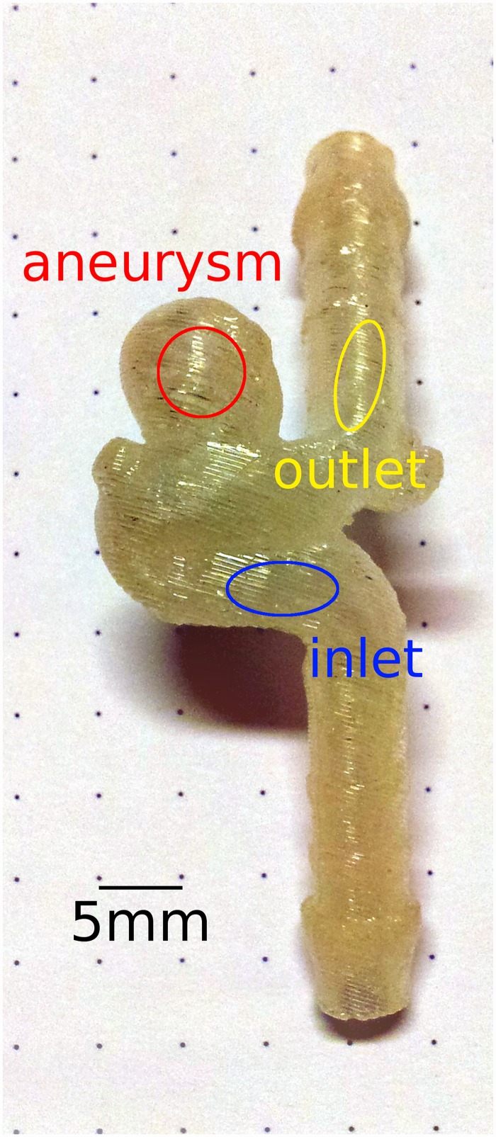
Materials and methods: The aneurysm model was of saccular morphology (7 mm dome height, 5 mm cross-section, 3-4 mm neck, 3.5 mm parent artery diameter) and connected to a peristaltic pump delivering a physiological flow (250 mL/min) and pulsation rate (70/min). High-resolution (4 h long) 4D phase contrast flow quantification (4D pc-fq) MRI was used to directly assess the hemodynamics of the model. Dynamic MPI, MRI, and DSA were performed with contrast agent injections (3 mL volume in 3 s) through a proximally placed catheter.
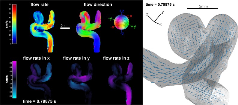
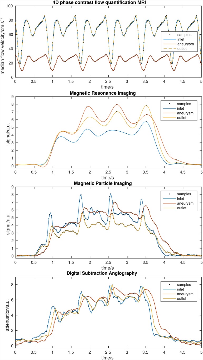
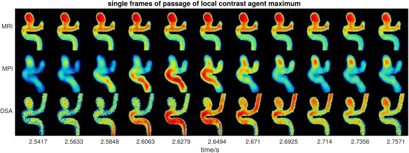
Results and discussion: 4D pc-fq measurements showed distinct pulsatile flow velocities (20-80 cm/s) as well as lower flow velocities and a vortex inside the aneurysm. All three dynamic methods (MPI, MRI, and DSA) also showed a clear pulsation pattern as well as delayed contrast agent dynamics within the aneurysm, which is most likely caused by the vortex within the aneurysm. Due to the high temporal resolution of MPI and DSA, it was possible to track the contrast agent bolus through the model and to estimate the average flow velocity (about 60 cm/s), which is in accordance with the 4D pc-fq measurements.
Conclusions: The ionizing radiation free, 4D high resolution MPI method is a very promising tool for imaging and characterization of hemodynamics in human. It carries the possibility of overcoming certain disadvantages of other modalities like considerably lower temporal resolution of dynamic MRI and limited 2D characteristics of DSA. Furthermore, additive manufacturing is the key for translating powerful pre-clinical techniques into the clinic.
Conflict of interest statement
Figures
References
-
- Knopp T, Weber A. Local System Matrix Compression for Efficient Reconstruction in Magnetic Particle Imaging. Adv Math Phys. 2015;2015: e472818 10.1155/2015/472818 - DOI
MeSH terms
Substances
LinkOut - more resources
Full Text Sources
Other Literature Sources
Medical
