

Smooth muscle cell sheet transplantation preserve cardiac function and minimize cardiac remodeling in a rat myocardial infarction model
- PMID: 27495170
- PMCID: PMC4974781
- DOI: 10.1186/s13019-016-0508-x
Smooth muscle cell sheet transplantation preserve cardiac function and minimize cardiac remodeling in a rat myocardial infarction model
Abstract
Background: We examined whether a vascular smooth muscle cell (SMC) sheet is effective in the treatment of a rat myocardial infarction (MI) model.
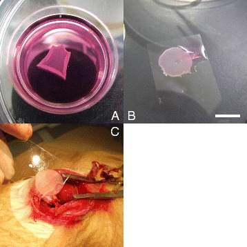
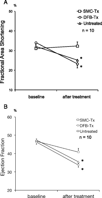
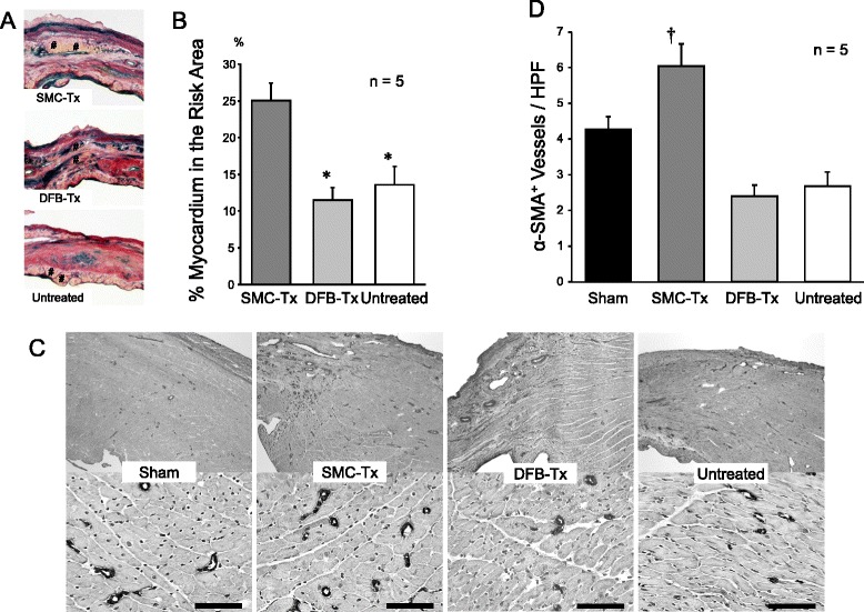
Methods: We examined the effect of SMC sheet on the cardiac function and cardiac remodeling in a rat MI model in comparison with their effect of dermal fibroblast (DFB) sheet in vivo. Furthermore, we estimated the apoptosis and secretion of angiogenic factor of SMC under hypoxic condition in comparison with DFB. Seven days after MI, monolayer cell sheets were transplanted on the infarcted area (SMC transplantation group, SMC-Tx; DFB transplantation group, DFB-Tx; no cell sheet transplantation group, Untreated; neither MI nor cell sheet transplantation group, Sham). We evaluated cardiac function by echocardiogram, degree of cardiac remodeling by histological examination, and secretion of angiogenic growth factor by enzyme immunoassay.
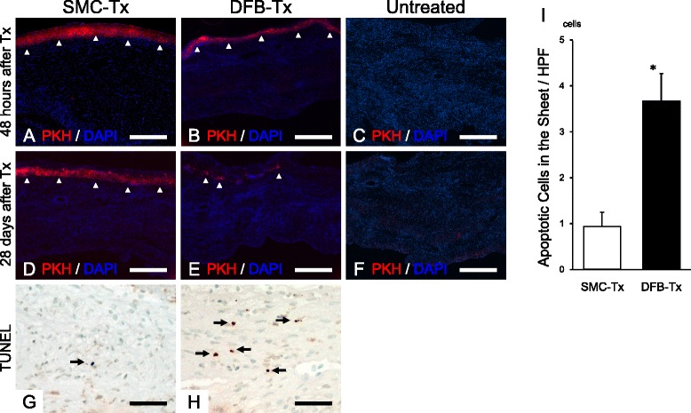
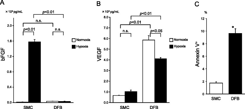
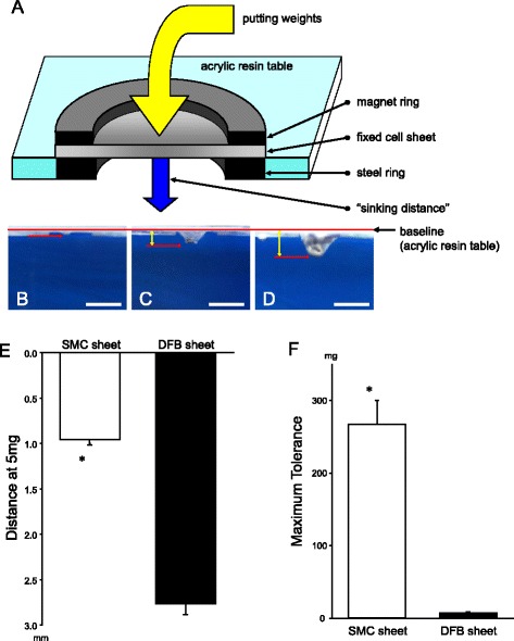
Results: Twenty-eight days after transplantation, SMC-Tx showed the following characteristics compared with the other groups: 1) significantly greater fractional area shortening (SMC-Tx, 32.3 ± 2.1 %; DFB-Tx, 23.3 ± 2.1 %; untreated, 25.1 ± 2.6 %), 2) suppressed left ventricular dilation, smaller scar expansion, and preserved wall thickness of the area at risk and the posterior wall, 3) decreased fibrosis, preserved myocardium in the scar area, and greater number of arterioles in border-zone, 4) tight attachment of SMC sheets on the scarred myocardium, and less apoptotic cell death. In in vitro experiments, SMCs secreted higher amounts of basic fibroblast growth factor (SMC, 157.7 ± 6.4 pg/ml; DFB, 3.1 ± 1.0 pg/ml), and showed less apoptotic cell death under hypoxia.
Conclusions: Our results illustrate that transplantation of SMC sheets inhibited the progression of cardiac remodeling and improve cardiac function. These beneficial effects may be due to superior SMC survival.
Keywords: Cell sheet; Cell survival; Myocardial infarction; Smooth muscle cells.
Figures
Similar articles
-
Alignment of inducible vascular progenitor cells on a micro-bundle scaffold improves cardiac repair following myocardial infarction.Basic Res Cardiol. 2017 Jul;112(4):41. doi: 10.1007/s00395-017-0631-4. Epub 2017 May 24. Basic Res Cardiol. 2017. PMID: 28540527
-
Layered smooth muscle cell-endothelial progenitor cell sheets derived from the bone marrow augment postinfarction ventricular function.J Thorac Cardiovasc Surg. 2017 Sep;154(3):955-963. doi: 10.1016/j.jtcvs.2017.04.081. Epub 2017 May 24. J Thorac Cardiovasc Surg. 2017. PMID: 28651946 Free PMC article.
-
p38 mitogen-activated protein kinase inhibition improves cardiac function and attenuates left ventricular remodeling following myocardial infarction in the rat.J Am Coll Cardiol. 2004 Oct 19;44(8):1679-89. doi: 10.1016/j.jacc.2004.07.038. J Am Coll Cardiol. 2004. PMID: 15489104
-
Tissue-engineered smooth muscle cell and endothelial progenitor cell bi-level cell sheets prevent progression of cardiac dysfunction, microvascular dysfunction, and interstitial fibrosis in a rodent model of type 1 diabetes-induced cardiomyopathy.Cardiovasc Diabetol. 2017 Nov 2;16(1):142. doi: 10.1186/s12933-017-0625-4. Cardiovasc Diabetol. 2017. PMID: 29096622 Free PMC article.
-
Clinical aspects of left ventricular diastolic function assessed by Doppler echocardiography following acute myocardial infarction.Dan Med Bull. 2001 Nov;48(4):199-210. Dan Med Bull. 2001. PMID: 11767125 Review.
Cited by
-
Three-Dimensional Human Cardiac Tissue Engineered by Centrifugation of Stacked Cell Sheets and Cross-Sectional Observation of Its Synchronous Beatings by Optical Coherence Tomography.Biomed Res Int. 2017;2017:5341702. doi: 10.1155/2017/5341702. Epub 2017 Feb 22. Biomed Res Int. 2017. PMID: 28326324 Free PMC article.
-
Heart regeneration with human pluripotent stem cells: Prospects and challenges.Bioact Mater. 2020 Jan 14;5(1):74-81. doi: 10.1016/j.bioactmat.2020.01.003. eCollection 2020 Mar. Bioact Mater. 2020. PMID: 31989061 Free PMC article. Review.
-
Role of smooth muscle cells in Cardiovascular Disease.Int J Biol Sci. 2020 Aug 21;16(14):2741-2751. doi: 10.7150/ijbs.49871. eCollection 2020. Int J Biol Sci. 2020. PMID: 33110393 Free PMC article. Review.
-
Promotion of right ventricular outflow tract reconstruction using a novel cardiac patch incorporated with hypoxia-pretreated urine-derived stem cells.Bioact Mater. 2021 Nov 30;14:206-218. doi: 10.1016/j.bioactmat.2021.11.021. eCollection 2022 Aug. Bioact Mater. 2021. PMID: 35310356 Free PMC article.
-
Human pluripotent stem cell-derived cardiac stromal cells and their applications in regenerative medicine.Stem Cell Res. 2020 May;45:101831. doi: 10.1016/j.scr.2020.101831. Epub 2020 Apr 27. Stem Cell Res. 2020. PMID: 32446219 Free PMC article. Review.
References
Publication types
MeSH terms
Substances
LinkOut - more resources
Full Text Sources
Other Literature Sources
Medical
