Review
. 2016 Aug;22(4 Movement Disorders):1208-26.
doi: 10.1212/CON.0000000000000362.

Ataxia

Review

Ataxia

Tetsuo Ashizawa et al. Continuum (Minneap Minn). 2016 Aug.

Abstract

Purpose of review: This article introduces the background and common etiologies of ataxia and provides a general approach to assessing and managing the patient with ataxia.

Recent findings: Ataxia is a manifestation of a variety of disease processes, and an underlying etiology needs to be investigated. Pure ataxia is rare in acquired ataxia disorders, and associated symptoms and signs almost always exist to suggest an underlying cause. While the spectrum of hereditary degenerative ataxias is expanding, special attention should be addressed to those treatable and reversible etiologies, especially potentially life-threatening causes. This article summarizes the diseases that can present with ataxia, with special attention given to diagnostically useful features. While emerging genetic tests are becoming increasingly available for hereditary ataxia, they cannot replace conventional diagnostic procedures in most patients with ataxia. Special consideration should be focused on clinical features when selecting a cost-effective diagnostic test.

Summary: Clinicians who evaluate patients with ataxia should be familiar with the disease spectrum that can present with ataxia. Following a detailed history and neurologic examination, proper diagnostic tests can be designed to confirm the clinical working diagnosis.


Figures
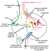
Figure 9-1
Figure 9-1
Afferent and efferent connections of the cerebellum. Main cerebellar afferent connections are by climbing fibers from the inferior olives through the inferior cerebellar peduncles, and pontine mossy fibers through the middle cerebellar peduncles. Cerebellar outputs are from the dentate nucleus and other deep cerebellar nuclei through the superior cerebellar peduncles.
Figure 9-2
Figure 9-2
Sagittal MRI of the patient in Case 9-1 showing atrophy of the cerebellum and brainstem. Reprinted with permission from Chakor RT, Bharote H, J Postgrad Med. © 2012 Journal of Postgraduate Medicine. http://www.jpgmonline.com/article.asp?issn=0022-3859;year=2012;volume=58;issue=4;spage=318;epage=325;aulast=Chakor.
Figure 9-3
Figure 9-3
Axial MRI of the patient in Case 9-2 showing a hot cross bun sign. Modified with permission from Srivastava T, et al, Neurology. © 2005 American Academy of Neurology. http://www.neurology.org/content/64/1/128.full.
Figure 9-4
Figure 9-4
Axial MRI of a 17-year-old girl with autosomal recessive spastic ataxia of Charlevoix-Saguenay showing degeneration of the corticospinal tract in the brainstem. The patient has had spasticity, ataxia, reduced fine motor function, and abnormal plantar responses since she was 5 years old. The patient’s family history was negative except for a cousin with spasticity. Consecutive axial T2-weighted MRI slices (A, B) demonstrate linear hypointensity in the pons. Reprinted with permission from Martin MH, et al, AJNR Am J Neuroradiol. © 2007 American Society of Neuroradiology. http://www.ajnr.org/content/28/8/1606.short.
Figure 9-5
Figure 9-5
Joubert syndrome. Axial MRI of a 5-year-old boy with delayed milestones, a prominent forehead and low-set ears, and cerebellar ataxia, hypotonia, and hyperreflexia, showing dysgenesis of the isthmus (the part of the brainstem between the pons and inferior colliculus), thick superior cerebellar peduncles, and hypoplasia of the vermis. These MRI findings produce the molar tooth sign. Reprinted with permission from Koshy B, et al, J Trop Pediatr. © 2009 The Authors. Published by Oxford University Press.
Figure 9-6
Figure 9-6
Fragile X tremor-ataxia syndrome. Axial T2-weighted MRI of a 76-year-old man showing increased signal intensity in the middle cerebellar peduncles. The patient presented to a movement disorders clinic with progressive gait ataxia since the age of 68, with later development of memory problems, occasional confusion, and tremor in his left hand during walking. His family history revealed developmental disability in his grandson through his daughter and premature ovarian failure in one of his granddaughters. Reprinted with permission from Hagerman PJ, Hagerman RJ, Nat Clin Pract Neurol. © 2007 Nature Publishing Group. http://www.nature.com/nrneurol/journal/v3/n2/full/ncpneuro0373.html.
Figure 9-7
Figure 9-7
Sensory ataxic neuropathy with dysarthria and ophthalmoparesis (with POLG mutation). Axial T2-weighted MRI of a 50-year-old man with a 4-year history of cerebellar ataxia, numb feet, diplopia, and slurred speech, showing well-defined symmetric signal abnormality in the cerebellar white matter, lateral to and sparing the dentate nuclei. Reprinted with permission from van Gaalen J, van de Warrenburg BP, Pract Neurol. © 2012, BMJ Publishing Group. pn.bmj.com/content/12/1/14.short.
Figure 9-8
Figure 9-8
Atrophy of the cervical spinal cord in a patient with Friedreich ataxia shown on T1-weighted sagittal section (A) and T2-weighted horizontal section (B) showing the atrophic cervical spinal cord (arrow) on MRI. Reprinted with permission from Mascalchi M, et al, AJR. © 1994 American Roentgen Ray Society. http://www.ajronline.org/doi/abs/10.2214/ajr.163.1.8010211.

References

    1. Németh AH, Kwasniewska AC, Lise S, et al. Next generation sequencing for molecular diagnosis of neurological disorders using ataxias as a model. Brain 2013;136(pt 10):3106–3118. doi:10.1093/brain/awt236. - PMC - PubMed
    1. Fogel BL, Lee H, Deignan JL, et al. Exome sequencing in the clinical diagnosis of sporadic or familial cerebellar ataxia. JAMA Neurol 2014;71(10):1237–1246. doi:10.1001/jamaneurol.2014.1944. - PMC - PubMed
    1. Barsottini OG, Albuquerque MV, Braga-Neto P, Pedroso JL. Adult onset sporadic ataxias: a diagnostic challenge. Arq Neuropsiquiatr 2014;72(3):232–240. doi:10.1590/0004-282X20130242. - PubMed
    1. Zeigelboim BS, Teive HA, Santos RS, et al. Audiological evaluation in spinocerebellar ataxia. Codas 2013;25(4):351–357. doi:10.1590/S2317-17822013005000001. - PubMed
    1. Whelan HT, Verma S, Guo Y, et al. Evaluation of the child with acute ataxia: a systematic review. Pediatr Neurol 2013;49(1):15–24. doi:10.1016/j.pediatrneurol.2012.12.005. - PubMed