. 2016 Aug 5;18(1):80.
doi: 10.1186/s13058-016-0741-1.

CUB domain-containing protein 1 and the epidermal growth factor receptor cooperate to induce cell detachment

Affiliations

CUB domain-containing protein 1 and the epidermal growth factor receptor cooperate to induce cell detachment

Mary E Law et al. Breast Cancer Res. .

Abstract

Background: While localized malignancies often respond to available therapies, most disseminated cancers are refractory. Novel approaches, therefore, are needed for the treatment of metastatic disease. CUB domain-containing protein1 (CDCP1) plays an important role in metastasis and drug resistance; the mechanism however, is poorly understood.

Methods: Breast cancer cell lines were engineered to stably express EGFR, CDCP1 or phosphorylation site mutants of CDCP1. These cell lines were used for immunoblot analysis or affinity purification followed by immunoblot analysis to assess protein phosphorylation and/or protein complex formation with CDCP1. Kinase activity was evaluated using phosphorylation site-specific antibodies and immunoblot analysis in in vitro kinase assays. Protein band excision and mass spectrometry was utilized to further identify proteins complexed with CDCP1 or ΔCDCP1, which is a mimetic of the cleaved form of CDCP1. Cell detachment was assessed using cell counting.

Results: This paper reports that CDCP1 forms ternary protein complexes with Src and EGFR, facilitating Src activation and Src-dependent EGFR transactivation. Importantly, we have discovered that a class of compounds termed Disulfide bond Disrupting Agents (DDAs) blocks CDCP1/EGFR/Src ternary complex formation and downstream signaling. CDCP1 and EGFR cooperate to induce detachment of breast cancer cells from the substratum and to disrupt adherens junctions. Analysis of CDCP1-containing complexes using proteomics techniques reveals that CDCP1 associates with several proteins involved in cell adhesion, including adherens junction and desmosomal cadherins, and cytoskeletal elements.

Conclusions: Together, these results suggest that CDCP1 may facilitate loss of adhesion by promoting activation of EGFR and Src at sites of cell-cell and cell-substratum contact.

Keywords: Adhesion; Breast cancer; CDCP1; E-cadherin; EGFR; Src.


Figures
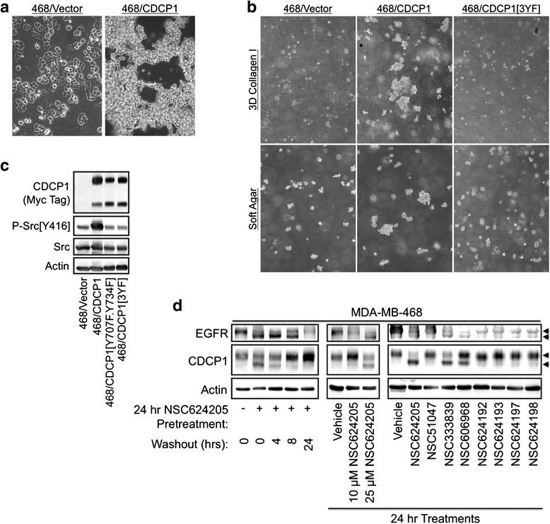
Fig. 1
Fig. 1
CDCP1 mediates suspension growth of MDA-MB-468 cells. a Morphology of the MDA-MB-468 vector control cell line or a line stably overexpressing wild-type CDCP1. CDCP1 induces suspension growth of cell cultures. b Comparison of MDA-MB-468 cell lines expressing wild-type CDCP1, or a phospho-defective form of CDCP1 in which tyrosine residues 707, 734, and 762 have been mutated to phenylalanine ([3YF]), grown in either soft agar or collagen I gels. Note that CDCP1-dependent suspension growth requires tyrosine phosphorylation. c Immunoblot analysis showing that the MDA-MB-468 cell lines stably express CDCP1 or its mutants at similar levels, but only wild-type CDCP1 increases activating Src phosphorylation above basal levels. d Treatment of MDA-MB-468 cells with Disulfide bond Disrupting Agents (DDAs) induces parallel mobility shifts of EGFR and CDCP1 in time- and concentration-dependent manners
Fig. 2
Fig. 2
CDCP1 forms ternary complexes with EGFR and Src. a Immunoprecipitation of FLAG-tagged CDCP1 from MDA-MB-468 and BT474 cells shows that CDCP1 associates with EGFR and HER2. b CDCP1 association with EGFR, Src, and PKCδ are significantly reduced either by mutation of Tyr734 to phenylalanine, or by a 24-hour treatment with 20 μM DDA NSC624203. c DDA treatment of cells dissociates EGFR/CDCP1 complexes in a concentration-dependent manner. d EGFR and Src form ternary complexes, as indicated by sequential CDCP1 and Src co-immunoprecipitation, and complex formation is reduced by treatment of the cells for 24 hours with 20 μM DDA NSC624203. e MDA-MB-468 cells transduced with adenoviruses encoding GFP, CDCP1 or PKCδ were stimulated with either 20 μM EGF or 100 μM sodium pervanadate for 20 minutes as indicated. Cell extracts were subjected to sequential anti-FLAG (CDCP1), anti-Src immunoprecipitation. Immunoprecipitates and the corresponding crude cell lysates were analyzed by immunoblot. f Lapatinib treatment (20 μM) for 24 hours reduces EGFR association with CDCP1. Dasatinib treatment (100 nM) for 24 hours does not alter EGFR association with CDCP1, but reduces overall tyrosine phosphorylation and EGFR phosphorylation on the Src site, Tyr845. In the MDA-MB-468 cell background, complex formation is not significantly altered by stimulation with 10 ng/ml EGF for 20 minutes. g Subjection of affinity purified CDCP1-containing complexes to in vitro kinase assays demonstrated ATP-dependent increases in overall tyrosine phosphorylation, EGFR phosphorylation on Tyr845 and Src phosphorylation on Tyr416. These phosphorylation events were blocked by the addition of 100 nM dasatinib to the kinase assays, but unaffected by 20 μM lapatinib
Fig. 3
Fig. 3
CDCP1 and EGFR cooperate to induce cell detachment from the substratum and to disrupt adherens junctions. a Immunoblot analysis of T47D cell lines stably expressing the indicated combinations of EGFR, CDCP1, and the Y734F mutant CDCP1. Coexpression of wild-type CDCP1 and EGFR results in a tyrosine-phosphorylated band that comigrates with EGFR. Similar results were observed whether the cells are plated on tissue culture plastic or type I collagen. b Coexpression of CDCP1 and EGFR result in enhanced EGF-dependent EGFR phosphorylation on Tyr845. This effect was blunted if wild-type CDCP1 was replaced with the Y734F mutant. c Treatment with DDA NSC624205 (20 μM) for 24 hours induces an electrophoretic mobility shift of EGFR, but not CDCP1, in the T47D cell background. d T47D cells stably expressing EGFR and CDCP1 were treated for 24 hours with vehicle or 20 μM DDA NSC333839 and stimulated for 20 minutes with or without 10 ng/ml EGF and analyzed by immunoblot. e The indicated cell lines were plated onto tissue culture plastic and treated with or without 10 ng/ml EGF for 24 hours and photographed. f Cells grown and treated as in Fig. 3b were gently pipetted with the culture medium and the detached suspension cells, and the cells remaining attached to the plates were counted. Results were plotted as the fraction of cells that were detached in triplicate determinations. Error bars represent standard deviation (SD). g Photomicrographs of the indicated cell lines treated as in Fig. 3e, but grown on the surface of collagen I gels. Note that EGFR and CDCP1 coexpression and EGF stimulation cooperate to induce suspension growth
Fig. 4
Fig. 4
CDCP1, EGFR, and EGF cooperate to induce E-cadherin internalization. a T47D cells stably expressing an E-cadherin-GFP fusion protein and either EGFR, CDCP1, or EGFR + CDCP1, or the corresponding vector control line were plated on glass coverslips coated with collagen I and treated for 24 hours with or without 10 ng/ml EGF. Intracellular E-cadherin-GFP localization was imaged by confocal immunofluorescence microscopy. b The indicated T47D cell lines were stimulated with or without 20 ng/ml EGF for 24 hours and subjected to cell surface biotinylation. Biotinylated proteins (external, Ext.) were isolated by streptavidin-agarose chromatography and unbound proteins were collected as non-biotinylated proteins (internal, Int.). Protein fractions were analyzed by immunoblot. c The indicated cell lines treated as in Fig. 4b were extracted with 1 % Triton X100 and analyzed by immunoblot
Fig. 5
Fig. 5
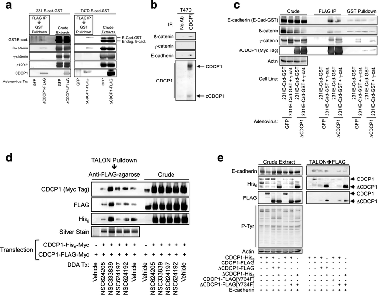
Characterization of E-cadherin/CDCP1-containing protein complexes. a MDA-MB-231 or T47D cell lines stably expressing an E-cadherin-glutathione S-transferase (E-cad-GST) fusion protein were transduced with an adenovirus encoding ΔCDCP1-FLAG or a control adenovirus encoding GFP. Cell extracts were subjected to sequential affinity purification using anti-FLAG agarose followed by glutathione-agarose. b T47D cell extracts were immunoprecipitated with a CDCP1 antibody and the immunoprecipitates were analyzed by immunoblot with the indicated antibodies. c The indicated stable cell lines were transduced with adenoviruses encoding GFP (control) or ΔCDCP1-FLAG and cell extracts were subjected to affinity purification with either anti-FLAG agarose or glutathione agarose. Affinity-purified proteins were analyzed by immunoblot. d HEK 293 cells were transiently transfected with the indicated tagged CDCP1 constructs and treated with the specified DDAs or vehicle. CDCP1 dimerization was monitored by TALON pulldown of His6-tagged proteins followed by purification with anti-FLAG agarose and immunoblot analysis of the sequentially affinity-purified material. e HEK 293 cells were transiently transfected with the indicated plasmid constructs and cell extracts were affinity purified sequentially using TALON resin and anti-FLAG agarose. The affinity-purified material was analyzed by immunoblot
Fig. 6
Fig. 6
Characterization of CDCP1-containing protein complexes reveals that MMP14 preferentially associates with cleaved CDCP1, while Galectin 1 only binds to full-length CDCP1. a Coomassie-stained SDS-PAGE gel of proteins affinity purified from MDA-MB-231 cells transduced with FLAG-tagged ΔCDCP1 or CDCP1 and treated for 15 minutes with or without the tyrosine phosphatase inhibitor sodium pervanadate (100 μM) to elevate overall tyrosine phosphorylation. Protein bands were excised and identified by mass spectrometry as described in the “Methods” section. b Extracts from MDA-MB-231 cells transduced with the indicated adenoviral vectors were affinity purified with anti-FLAG agarose to isolate CDCP1-containing complexes and the purified material or corresponding crude lysates were analyzed by immunoblot. c Study performed as in Fig. 5b except that cells were treated for 24 hours with either vehicle, 20 μM of the matrix metalloproteinase inhibitor GM6001 or 100 nM of the Src-family kinase inhibitor dasatinib. d Silver-stained SDS-PAGE gel of proteins isolated by anti-FLAG affinity purification from MDA-MB-231 cells transduced with the indicated adenoviruses and treated for 15 minutes with or without 100 μM sodium pervanadate. A low-molecular-mass protein was observed (red arrow) that was present when full-length forms of CDCP1 were expressed, but not when cleaved forms of CDCP1 (ΔCDCP1) were expressed. e HEK 293 cells were transiently transfected with the indicated vectors and CDCP1 or ΔCDCP1-containing complexes were isolated with anti-FLAG agarose and analyzed by immunoblot
Fig. 7
Fig. 7
CDCP1 complexes are similar between breast and pancreatic cancer cell lines and are stable in a variety of detergents. a MDA-MB-231 or T47D cells were transduced with the indicated adenoviral vectors, CDCP1-containing complexes were isolated with anti-FLAG agarose, and the presence of components of adherens and desmosomal junctions was assessed by immunoblot analysis. b This study was carried out similarly to the study in Fig. 6a, except that the pancreatic cancer cell lines AsPC1 or PANC1 were used. c AsPC1 cells were transduced with control (GFP) or ΔCDCP1 adenoviruses, the cells were extracted with the indicated detergents, and ΔCDCP1-associated proteins were examined by immunoblot (left panel) or total protein levels were assessed by Coomassie staining of the same samples resolved by SDS-PAGE (right panel)
Fig. 8
Fig. 8
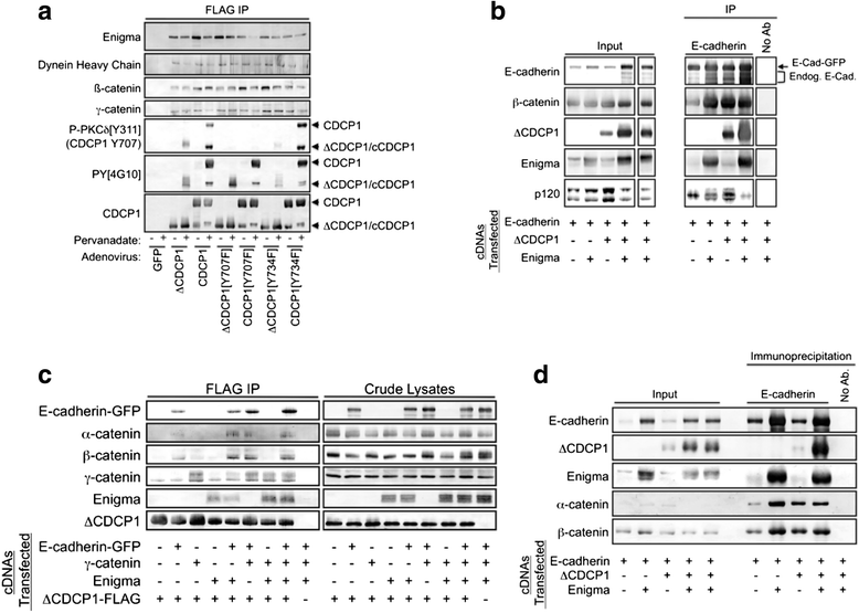
Enigma/PDLIM7 as a novel CDCP1-associated protein. a MDA-MB-231 cells were transduced with the indicated recombinant adenoviruses, then treated for 15 minutes with 100 μM sodium pervanadate just before cell lysates were prepared. The various forms of CDCP1 were isolated by anti-FLAG affinity chromatography and CDCP1-associated proteins were identified by immunoblot. Note that the phospho-PKCδ[Y311] antibody cross-reacts with phosphorylated Tyr707 of CDCP1 and that staining was absent when the Y707F mutant of CDCP1 was employed. b HEK 293 cells were transfected with the indicated constructs, cell lysates were immunoprecipitated with an E-cadherin antibody, and the immunoprecipitates were analyzed by immunoblot. c HEK 293 cells were transfected with the indicated constructs, cell lysates were immunoprecipitated with anti-FLAG agarose, and the immunoprecipitates were analyzed by immunoblot. d HEK 293 cells were transfected with the indicated constructs, cell lysates were immunoprecipitated with an E-cadherin antibody, and the immunoprecipitates were analyzed by immunoblot
Fig. 9
Fig. 9
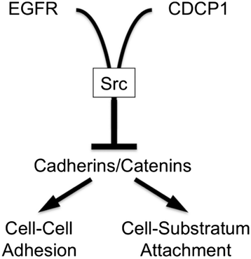
Model for CDCP1 influences on cell-cell and substratum adhesion through a variety of mechanisms. We propose that CDCP1 participates in complexes with EGFR and Src leading to Src activation and EGFR transactivation, and that CDCP1 also participates in complexes that contain cell adhesion proteins such as cadherins and catenins. EGFR and Src have been established to reduce E-cadherin-mediated cell-cell adhesion and to induce E-cadherin internalization into cytoplasmic vesicles. Therefore, CDCP1 may abrogate cadherin adhesive function through the assembly of “anti-adhesive” complexes

References

    1. Brown TA, Yang TM, Zaitsevskaia T, Xia Y, Dunn CA, Sigle RO, Knudsen B, Carter WG. Adhesion or plasmin regulates tyrosine phosphorylation of a novel membrane glycoprotein p80/gp140/CUB domain-containing protein 1 in epithelia. J Biol Chem. 2004;279:14772–83. doi: 10.1074/jbc.M309678200. - DOI - PubMed
    1. Scherl-Mostageer M, Sommergruber W, Abseher R, Hauptmann R, Ambros P, Schweifer N. Identification of a novel gene, CDCP1, overexpressed in human colorectal cancer. Oncogene. 2001;20:4402–8. doi: 10.1038/sj.onc.1204566. - DOI - PubMed
    1. Bhatt AS, Erdjument-Bromage H, Tempst P, Craik CS, Moasser MM. Adhesion signaling by a novel mitotic substrate of src kinases. Oncogene. 2005;24:5333–43. doi: 10.1038/sj.onc.1208582. - DOI - PMC - PubMed
    1. Miura S, Hamada S, Masamune A, Satoh K, Shimosegawa T. CUB-domain containing protein 1 represses the epithelial phenotype of pancreatic cancer cells. Exp Cell Res. 2014;321:209–18. doi: 10.1016/j.yexcr.2013.12.019. - DOI - PubMed
    1. Gioia R, Leroy C, Drullion C, Lagarde V, Etienne G, Dulucq S, Lippert E, Roche S, Mahon FX, Pasquet JM. Quantitative phosphoproteomics revealed interplay between Syk and Lyn in the resistance to nilotinib in chronic myeloid leukemia cells. Blood. 2011;118:2211–21. doi: 10.1182/blood-2010-10-313692. - DOI - PubMed