

Knockout maternal adiponectin increases fetal growth in mice: potential role for trophoblast IGFBP-1
- PMID: 27495989
- PMCID: PMC5042853
- DOI: 10.1007/s00125-016-4061-x
Knockout maternal adiponectin increases fetal growth in mice: potential role for trophoblast IGFBP-1
Abstract
Aims/hypothesis: The main objective of this study was to investigate whether maternal adiponectin regulates fetal growth through the endocrine system in the fetal compartment.
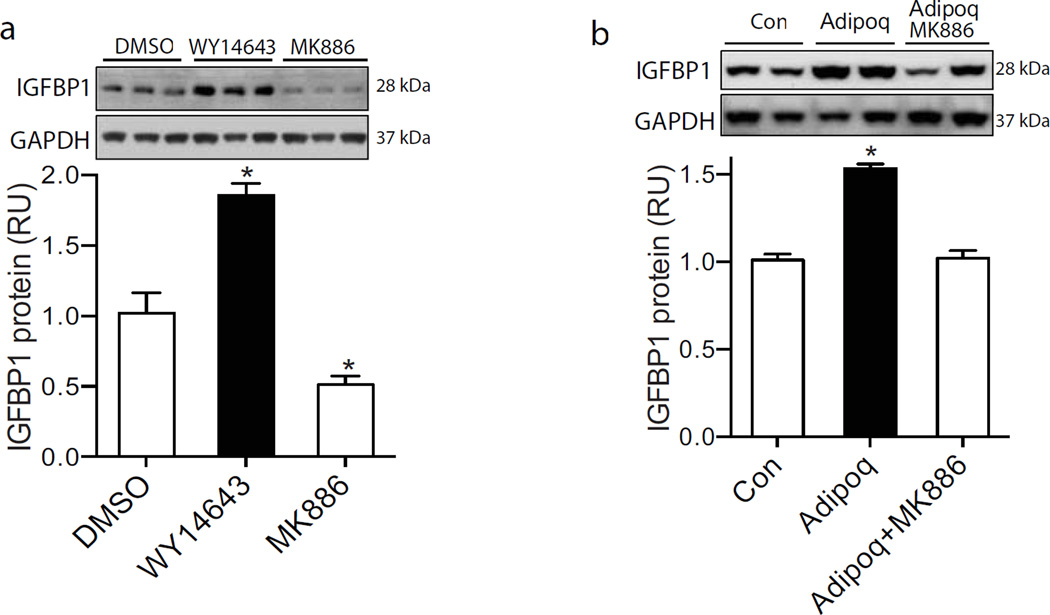
Methods: Adiponectin knockout (Adipoq (-/-) ) mice and in vivo adenovirus-mediated reconstitution were used to study the regulatory effect of maternal adiponectin on fetal growth. Primary human trophoblast cells were treated with adiponectin and a specific peroxisome proliferator-activated receptor α (PPARα) agonist or antagonist to study the underlying mechanism through which adiponectin regulates fetal growth.
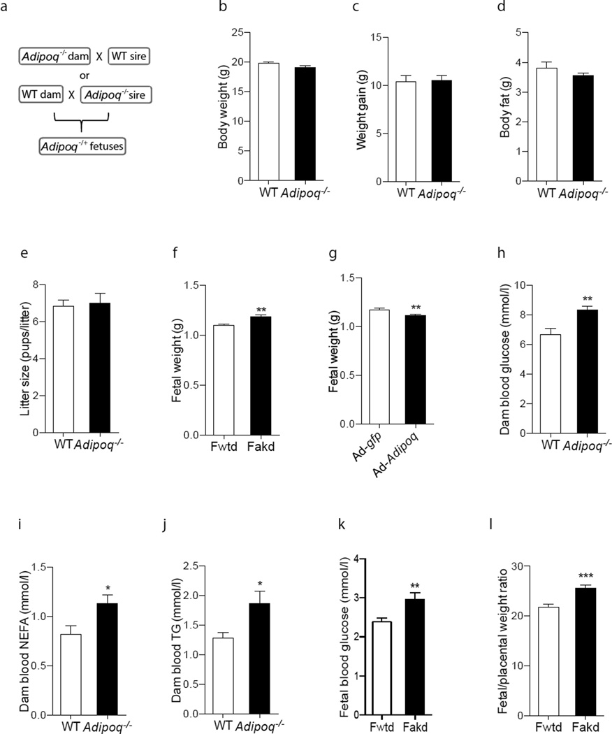
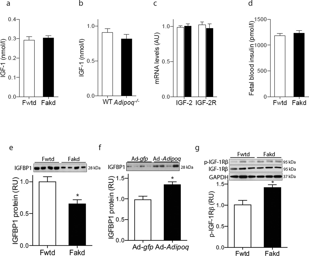
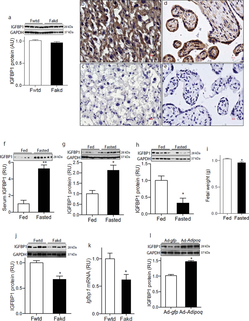
Results: The body weight of fetuses from Adipoq (-/-) dams was significantly greater than that of wild-type dams at both embryonic day (E)14.5 and E18.5. Adenoviral vector-mediated maternal adiponectin reconstitution attenuated the increased fetal body weight induced by maternal adiponectin deficiency. Significantly increased blood glucose, triacylglycerol and NEFA levels were observed in Adipoq (-/-) dams, suggesting that nutrient supply contributes to maternal adiponectin-regulated fetal growth. Although fetal blood IGF-1 concentrations were comparable in fetuses from Adipoq (-/-) and wild-type dams, remarkably low levels of IGF-binding protein 1 (IGFBP-1) were observed in the serum of fetuses from Adipoq (-/-) dams. IGFBP-1 was identified in the trophoblast cells of human and mouse placentas. Maternal fasting robustly increased IGFBP-1 levels in mouse placentas, while reducing fetal weight. Significantly low IGFBP-1 levels were found in placentas of Adipoq (-/-) dams. Adiponectin treatment increased IGFBP-1 levels in primary cultured human trophoblast cells, while the PPARα antagonist, MK886, abolished this stimulatory effect.
Conclusions/interpretation: These results indicate that, in addition to nutrient supply, maternal adiponectin inhibits fetal growth by increasing IGFBP-1 expression in trophoblast cells.
Keywords: Adiponectin; Fetus; Growth; IGF-binding protein; Placenta.
Figures
Similar articles
-
Adiponectin Deficiency Impairs Maternal Metabolic Adaptation to Pregnancy in Mice.Diabetes. 2017 May;66(5):1126-1135. doi: 10.2337/db16-1096. Epub 2017 Jan 10. Diabetes. 2017. PMID: 28073830 Free PMC article.
-
Chronic maternal infusion of full-length adiponectin in pregnant mice down-regulates placental amino acid transporter activity and expression and decreases fetal growth.J Physiol. 2012 Mar 15;590(6):1495-509. doi: 10.1113/jphysiol.2011.226399. Epub 2012 Jan 30. J Physiol. 2012. PMID: 22289908 Free PMC article.
-
Adiponectin Promotes Maternal β-Cell Expansion Through Placental Lactogen Expression.Diabetes. 2021 Jan;70(1):132-142. doi: 10.2337/db20-0471. Epub 2020 Oct 21. Diabetes. 2021. PMID: 33087456 Free PMC article.
-
Review: Adiponectin--the missing link between maternal adiposity, placental transport and fetal growth?Placenta. 2013 Mar;34 Suppl:S40-5. doi: 10.1016/j.placenta.2012.11.024. Epub 2012 Dec 13. Placenta. 2013. PMID: 23245987 Free PMC article. Review.
-
Spatial and temporal patterns of expression of messenger RNA for insulin-like growth factors and their binding proteins in the placenta of man and laboratory animals.Placenta. 2000 May;21(4):289-305. doi: 10.1053/plac.1999.0498. Placenta. 2000. PMID: 10833363 Review.
Cited by
-
Placental RNA sequencing implicates IGFBP1 in insulin sensitivity during pregnancy and in gestational diabetes.Res Sq [Preprint]. 2023 Oct 27:rs.3.rs-3464151. doi: 10.21203/rs.3.rs-3464151/v1. Res Sq. 2023. Update in: Nat Med. 2024 Jun;30(6):1689-1695. doi: 10.1038/s41591-024-02936-5. PMID: 37961187 Free PMC article. Updated. Preprint.
-
The Relationship between Maternal Plasma Leptin and Adiponectin Concentrations and Newborn Adiposity.Nutrients. 2017 Feb 23;9(3):182. doi: 10.3390/nu9030182. Nutrients. 2017. PMID: 28241462 Free PMC article.
-
Placental function in maternal obesity.Clin Sci (Lond). 2020 Apr 30;134(8):961-984. doi: 10.1042/CS20190266. Clin Sci (Lond). 2020. PMID: 32313958 Free PMC article. Review.
-
The gonadal expression pattern of lipocalin‑2 and 24p3 receptor is modified in the gonads of the offspring of obese rats.Mol Med Rep. 2020 Aug;22(2):1409-1419. doi: 10.3892/mmr.2020.11226. Epub 2020 Jun 12. Mol Med Rep. 2020. PMID: 32627017 Free PMC article.
-
Trophoblast-specific overexpression of adiponectin receptor 2 causes fetal growth restriction in pregnant mice.FASEB J. 2024 Oct 15;38(19):e70100. doi: 10.1096/fj.202302143R. FASEB J. 2024. PMID: 39387608
References
-
- Ogden CL, Fryar CD, Flegal KM. National Center for Health Statistics. Hyattsville, MD: National Center for Health Statistics; 2015. Prevalence of obesity among adults and youth: United States, 2011–2014. - PubMed
-
- Oken E. Fetal origins of obesity. Obesity research. 2003;11:496–506. - PubMed
-
- Rooney K, Ozanne SE. Maternal over-nutrition and offspring obesity predisposition: targets for preventative interventions. Int J Obes (Lond) 2011;35:883–890. - PubMed
-
- Freinkel N. Banting Lecture 1980. Of pregnancy and progeny. Diabetes. 1980;29:1023–1035. - PubMed
-
- Barker DJ, Thornburg KL. The obstetric origins of health for a lifetime. Clin Obstet Gynecol. 2013;56:511–519. - PubMed
Publication types
MeSH terms
Substances
Grants and funding
LinkOut - more resources
Full Text Sources
Other Literature Sources
Molecular Biology Databases
Research Materials
Miscellaneous
